Comparative Study
. 2006 Sep 15;174(6):689-98.
doi: 10.1164/rccm.200502-276OC. Epub 2006 Jun 1.

Neutrophil cytoskeletal rearrangements during capillary sequestration in bacterial pneumonia in rats

Affiliations
Comparative Study

Neutrophil cytoskeletal rearrangements during capillary sequestration in bacterial pneumonia in rats

Kazuo Yoshida et al. Am J Respir Crit Care Med. .

Abstract

Rationale: Neutrophils accumulate in pulmonary capillaries during acute inflammation. Initial events in injury recognition and sequestration do not occur through selectin-mediated rolling. Cytoskeletal rearrangements, as assessed by submembrane F-actin rims, result in poorly deformable neutrophils that may not pass through capillaries.

Objective: To test the hypothesis that neutrophils sequestering during pneumonia contain F-actin rims and to determine the roles of CD11/CD18, L-selectin expression, and neutrophil-platelet adhesion in neutrophil sequestration.

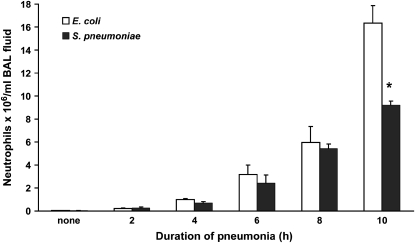
Methods: Neutrophils were compared in blood obtained simultaneously from venous and arterial sites before and 4 h after instillation of Streptococcus pneumoniae or Escherichia coli in rats.

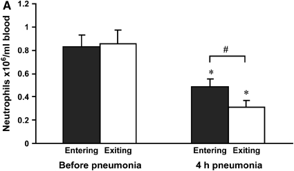
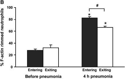
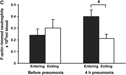
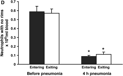
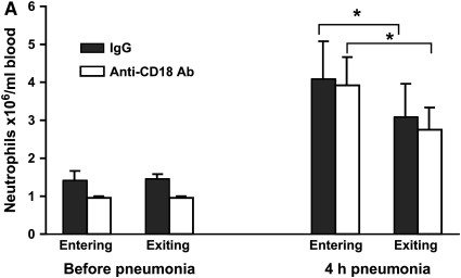
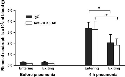
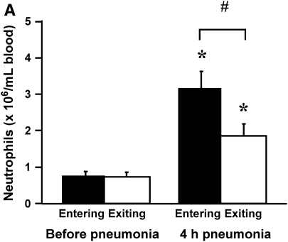
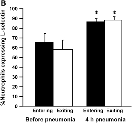
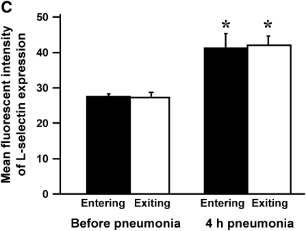
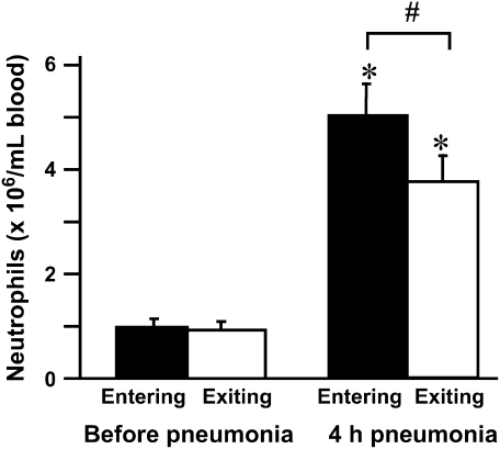
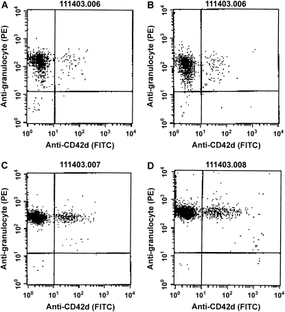
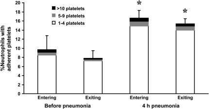
Measurements and main results: At 4 h of pneumonia, the number of neutrophils was greater in the venous blood entering the lungs than in the arterial blood leaving the lungs, indicating that neutrophil sequestration was occurring. More neutrophils entering the lungs contained F-actin rims than did neutrophils exiting, and the venous-arterial difference in F-actin-rimmed neutrophil counts completely accounted for sequestration. In E. coli pneumonia, in which neutrophil adhesion is mediated by CD11/CD18, CD18 blockade 15 min before blood samples were obtained did not prevent this sequestration of F-actin-rimmed neutrophils. Neutrophils expressing high or low levels of L-selectin or of neutrophils that bound platelets while circulating did not preferentially sequester.

Conclusions: Neutrophils with cytoskeletal rearrangements preferentially sequester within the lungs during pneumonia, and this sequestration is not due to CD11/CD18-mediated adhesion, L-selectin expression, or platelet adhesion to neutrophils, suggesting that cytoskeletal rearrangements result in sequestration of neutrophils.


Figures

<b>Figure 1.</b>
Figure 1.
Neutrophil emigration into airspaces of lungs. Neutrophil migration had initiated by 4 h after instillation of either E. coli or S. pneumoniae. Neutrophils in bronchoalveolar lavage (BAL) fluid were similar in number at all time points except 10 h after instillation, when the recruitment induced by E. coli was greater than that induced by S. pneumoniae. Neutrophil migration increased 8- to 15-fold between 4 and 10 h. n = 5 at each time point except 10 h, where n = 2. *Significant difference between E. coli and S. pneumoniae, p < 0.05.
<b>Figure 2.</b>
Figure 2.
(A) Neutrophil counts in blood samples drawn simultaneously from the venous (entering the lungs) and arterial (exiting the lungs) circulation. Venous and arterial neutrophil counts were similar before pneumonia. Four hours after initiation of S. pneumoniae pneumonia, both venous and arterial counts were less than before pneumonia. The count was less in blood exiting the lungs compared with that entering the lungs, indicating that neutrophil sequestration within the lungs was occurring. (B) Percentage of circulating neutrophils that contained F-actin rims in S. pneumoniae pneumonia. The percentage of circulating neutrophils with F-actin rims increased by 4 h after instillation of organisms. At 4 h, the percentage of F-actin–rimmed neutrophils was less in venous blood than in arterial blood. (C) Number of F-actin–rimmed neutrophils in venous and arterial blood samples before and during S. pneumoniae pneumonia. The numbers of F-actin–rimmed neutrophils in venous blood and arterial blood were similar before pneumonia. Four hours after initiation of pneumonias, fewer F-actin–rimmed neutrophils were present in the blood exiting than entering the lungs, indicating that neutrophils containing F-actin rims sequestered in the lungs. (D) Number of neutrophils without F-actin rims in S. pneumoniae pneumonia. Neutrophils without rims were similar in number in venous and arterial blood at both time points. *Significantly less than the value determined at the same site before pneumonia, p < 0.05. #Significantly less than the value in the venous blood entering the lungs, p < 0.05.
<b>Figure 3.</b>
Figure 3.
Circulating neutrophil counts (A) and F-actin–rimmed neutrophil counts (B) in E. coli pneumonia. Neutrophil counts significantly increased in both venous and arterial blood samples compared with the counts in blood samples obtained before pneumonia. The circulating counts at 4 h were lower in arterial blood (exiting the lungs) than in venous blood (entering the lungs) of rats given nonimmune IgG. When rats were pretreated with a blocking anti-CD18 antibody given intravenously 15 min before blood samples were obtained, a similar difference between venous and arterial neutrophil counts was observed, as seen in animals given IgG. E. coli also induced an increase in the number of circulating F-actin–rimmed neutrophils by 4 h. The number of F-actin–rimmed neutrophils was less in arterial than in venous blood. Blockade of CD18 did not prevent this difference, indicating that CD11/CD18 is not required for the sequestration of F-actin–rimmed neutrophils within the pulmonary microvasculature. *Significantly less than counts in the venous blood entering the lungs, p < 0.05.
<b>Figure 4.</b>
Figure 4.
(A) The number of circulating neutrophils, (B) the percentage of neutrophils that expressed L-selectin, and (C) the mean fluorescence intensity and standard deviation of L-selectin on neutrophils obtained from venous and arterial blood samples before and 4 h after instillation of E. coli. Similar to the rats studied in Figures 2A and 3A, E. coli induced a significant increase in circulating neutrophil counts by 4 h (A). Arterial neutrophil counts were significantly less than venous counts, indicating that sequestration was ongoing at this time. The percentage of neutrophils expressing L-selectin was significantly increased at 4 h after pneumonia (B). Neutrophils from venous and arterial blood samples were similar. The mean fluorescence intensity of L-selectin labeling and the standard deviation increased in animals with 4-h pneumonias (C). There was no difference in venous compared with arterial values. Similarly, standard deviations increased at 4 h after pneumonia, and were similar between venous and arterial samples. *Significantly greater than neutrophil counts before pneumonia, p < 0.05. #Significantly greater than the value obtained in arterial blood samples, p < 0.05.
<b>Figure 5.</b>
Figure 5.
Circulating neutrophil counts in rats with E. coli pneumonia. As demonstrated in Figures 3A and 4A, neutrophil counts increased in rats after instillation of E. coli pneumonia, and the arterial counts were less than the venous counts, indicating that sequestration of neutrophils was occurring in the lungs. In these samples, the percentage of neutrophils to which platelets were bound is shown in Figures 6 and 7. *Significantly greater than the number before pneumonia, p < 0.05. #Significantly less than the venous counts, p < 0.05.
<b>Figure 6.</b>
Figure 6.
An example of data obtained by flow cytometry from a single rat, identifying the percentage of neutrophils to which platelets were bound in blood samples. Blood samples were obtained before pneumonia from venous (A) and arterial (B) catheters and 4 h after instillation of E. coli, also from venous (C) and arterial (D) sites. Cells were labeled with an anti-granulocyte antibody (clone HIS48) followed by phycoerythrin-labeled anti-mouse IgM and a fluorescein isothiocyanate–labeled mouse anti-rat CD42d monoclonal antibody (clone RPM-4) as described in Methods. Flow cytometry was performed by gating on the neutrophils and determining the number of HIS48-positive cells that expressed the platelet marker CD42d (upper right region of each scattergram).
<b>Figure 7.</b>
Figure 7.
The percentage of circulating neutrophils that bound platelets in the venous and arterial blood before and at 4 h after instillation of E. coli. This percentage increased approximately two-fold during E. coli pneumonia, and this change was due to an increase in the percentage of neutrophils binding 1–4 platelets. No increase was observed in the percentage of neutrophils binding 5–9 or more than 10 platelets. This increase occurred in both venous and arterial blood samples. There was no significant difference in the percentage of neutrophils that bound platelets in the venous compared to the arterial blood samples before pneumonia or at 4 h after instillation. * Significantly greater than the percentage of neutrophils that bound platelets before pneumonia, p < 0.05.

References

    1. Loosli CG, Baker RF. Acute experimental pneumonococcal (type I) pneumonia in the mouse: the migration of leukocytes from the pulmonary capillaries into the alveolar spaces as revealed by the electron microscope. Trans Am Clin Climatol Assoc 1962;74:15–28. - PMC - PubMed
    1. Shaw JO. Leukocytes in chemotactic fragment–induced lung inflammation. Am J Pathol 1980;101:283–302. - PMC - PubMed
    1. Walker DC, Behzad AR, Chu F. Neutrophil migration through preexisting holes in the basal laminae of alveolar capillaries and epithelium during streptococcal pneumonia. Microvasc Res 1995;50:397–416. - PubMed
    1. Behzad AR, Chu F, Walker DC. Fibroblasts are in a position to provide directional information to migrating neutrophils during pneumonia in rabbit lungs. Microvasc Res 1996;51:303–316. - PubMed
    1. Downey GP, Worthen GS, Henson PM, Hyde DM. Neutrophil sequestration and migration in localized pulmonary inflammation: capillary localization and migration across the interalveolar septum. Am Rev Respir Dis 1993;147:168–176. - PubMed

Publication types

MeSH terms