

Increased number of subcortical hyperintensities on MRI in children and adolescents with Tourette's syndrome, obsessive-compulsive disorder, and attention deficit hyperactivity disorder
- PMID: 16741215
- PMCID: PMC2367225
- DOI: 10.1176/ajp.2006.163.6.1106
Increased number of subcortical hyperintensities on MRI in children and adolescents with Tourette's syndrome, obsessive-compulsive disorder, and attention deficit hyperactivity disorder
Abstract
Objective: To investigate whether cerebral hyperintensities on T2-weighted magnetic resonance images (MRI) are associated with childhood neuropsychiatric disorders.
Method: The authors compared the frequency of cortical and subcortical cerebral hyperintensities in 100 children and adolescents with Tourette's syndrome, obsessive-compulsive disorder (OCD), or attention deficit hyperactivity disorder (ADHD) and 32 healthy comparison subjects.
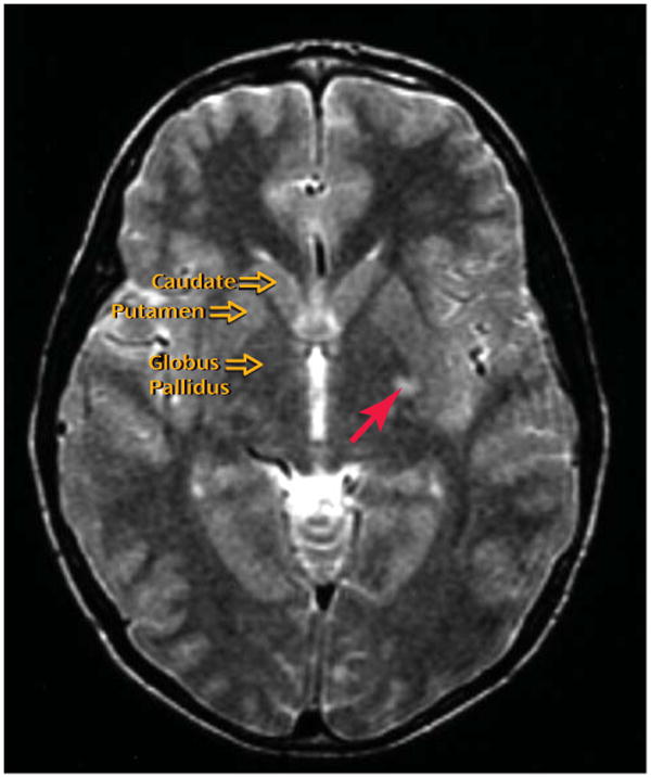
Results: The frequency of cerebral hyperintensities was significantly higher in subjects with Tourette's syndrome, OCD, or ADHD than in healthy comparison subjects; each diagnostic group seemed to contribute to this effect. Among the patient groups, the likelihood of detecting cerebral hyperintensities in the subcortex (primarily the basal ganglia and thalamus) was significantly greater than in the cortex.
Conclusions: A childhood diagnosis of Tourette's syndrome, OCD, or ADHD significantly increased the likelihood of detecting cerebral hyperintensities, particularly in the subcortex, supporting the notion that subcortical injury may play a role in the pathophysiology of these conditions.
Figures
References
-
- Mantyla R, Aronen HJ, Salonen O, Pohjasvaara T, Korpelainen M, Peltonen T, Standertskjold-Nordenstam, Kaste M, Erkinjuntti T. MRI white matter hyperintensities and mechanism of ischemic stroke. Stroke. 1999;30(10):2053–2058. - PubMed
-
- Pomper MG, Miller TJ, Stone JH, Tidmore WC, Hellman DB. CNS vasculitis in autoimmune disease. Am J Neuroradiol. 1999;20:75–85. - PubMed
-
- Giedd JN, Rapoport JL, Kruesi MJ, Parker C, Schapiro MB, Allen AJ, Leonard HL, Kaysen D, Dickstein DP, Marsh WL, Kozuch PL, Vaituzis AC, Hamburger SD, Swedo SE. Sydenham's chorea magnetic resonance imaging of the basal ganglia. Neurology. 1995;45:2199–2202. - PubMed
-
- Bodner SM, Peterson BS. Pediatric autoimmune neuropsychiatric disorders associated with streptococcus. Dir Psychiatry. 2003;23:235–251.
Publication types
MeSH terms
Grants and funding
LinkOut - more resources
Full Text Sources
Medical
