

Noninvasive measurement of cardiovascular function in mice with high-temporal-resolution small-animal PET
- PMID: 16741307
- PMCID: PMC4348007
Noninvasive measurement of cardiovascular function in mice with high-temporal-resolution small-animal PET
Abstract
The aim of this study was to explore the feasibility of determining parameters of cardiovascular function in mice noninvasively by high-temporal-resolution imaging with a dedicated small-animal PET system.
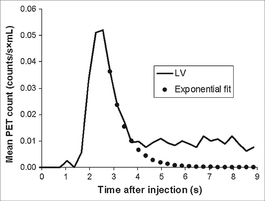
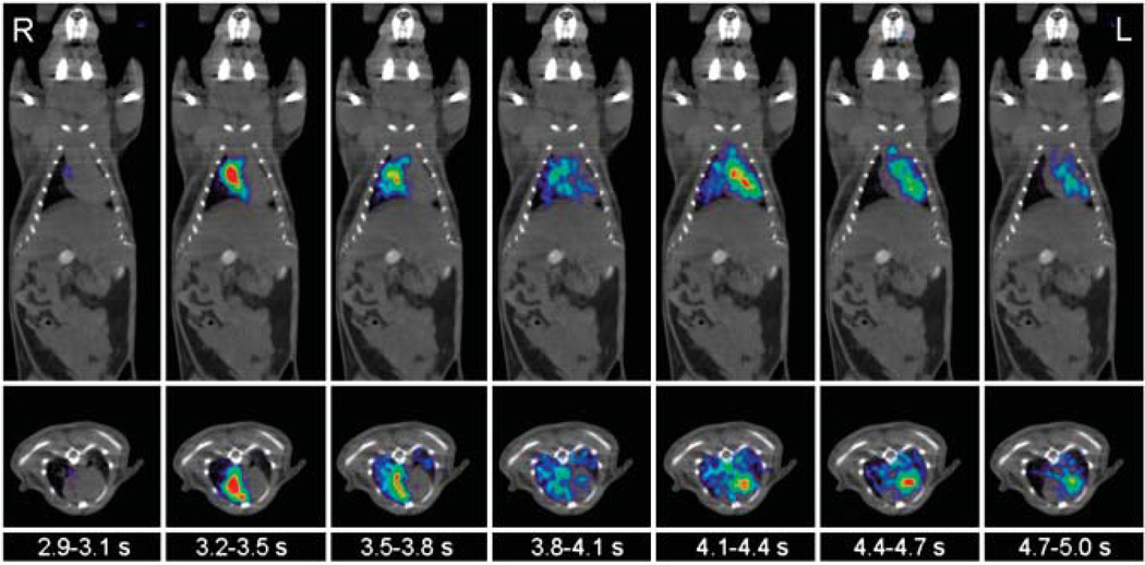
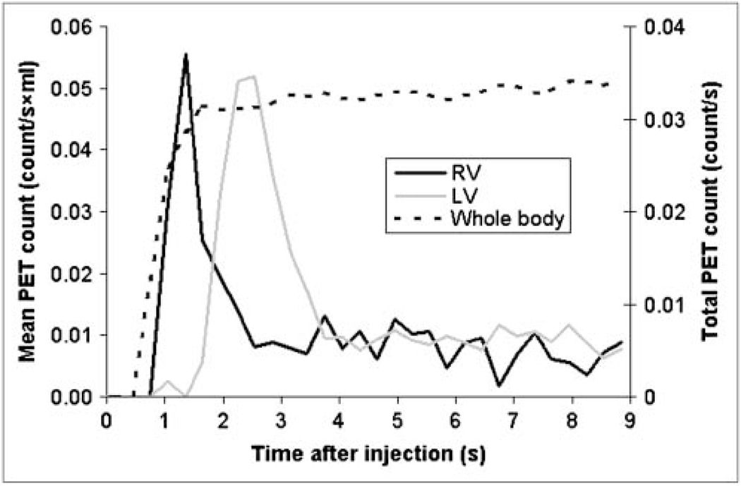
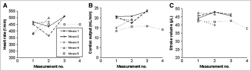
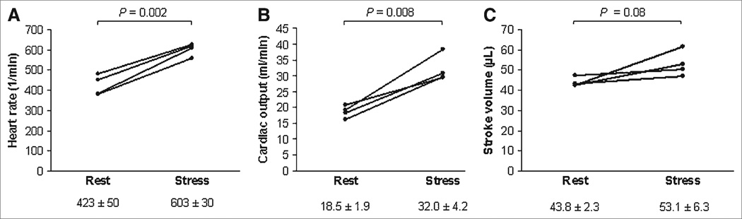
Methods: Twenty-five anesthetized mice (28.8 +/- 4.6 g) were injected via an intravenous catheter with a 30-microL bolus of (18)F-FDG (8-44 MBq). The first 9 s of data were reconstructed into 30 frames of 0.3 s using filtered backprojection. The time-activity curve derived from a left ventricle volume of interest was corrected for tracer recirculation and partial volume. Cardiac output was calculated by the Stewart-Hamilton method, in which cardiac output is total injected activity divided by the area under the left ventricle time-activity curve. Cardiac output divided by body weight was defined as cardiac index; cardiac output divided by heart rate yielded the stroke volume. In 5 mice, measurements were repeated 2-4 times to assess reproducibility. In 4 mice, the hemodynamic response to dobutamine was examined by measuring heart rate, cardiac output, and stroke volume.
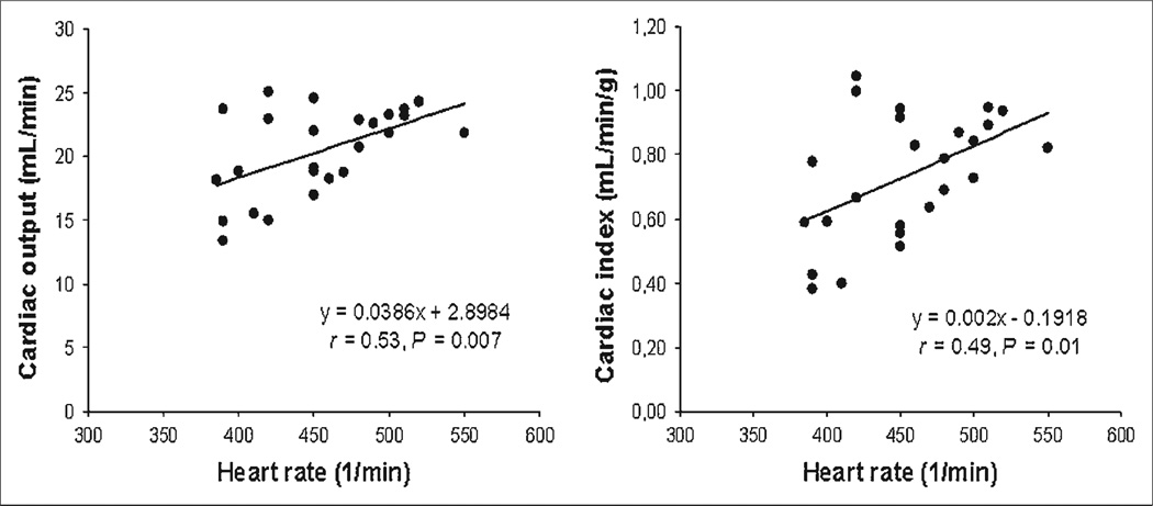
Results: The cardiac output averaged 20.4 +/- 3.4 mL/min; in the repeated measurements, the parameter displayed a mean percentage SD per mouse of 10% +/- 6%. The cardiac index averaged 0.73 +/- 0.19 mL/min/g and the stroke volume 45.0 +/- 6.9 microL, and both correlated with heart rate (r = 0.53, P = 0.007, and r = 0.49, P = 0.01, respectively). During dobutamine stress, heart rate increased from 423 +/- 50 to 603 +/- 30 beats/min (P = 0.002) and cardiac output increased from 18.5 +/- 1.9 to 32.0 +/- 4.2 mL/min (P = 0.008).
Conclusion: Parameters of cardiovascular function can be measured in mice noninvasively by radionuclide angiography using high-temporal-resolution small-animal PET. Measured values of cardiac output and stroke volume are reproducible and comparable to those obtained with MRI. The approach permits the monitoring of changes in cardiovascular function in response to pharmacologic intervention.
Figures
References
-
- Cherry SR, Gambhir SS. Use of positron emission tomography in animal research. ILAR J. 2001;42:219–232. - PubMed
-
- Gambhir SS, Barrio JR, Herschman HR, Phelps ME. Assays for noninvasive imaging of reporter gene expression. Nucl Med Biol. 1999;26:481–490. - PubMed
-
- Sen L, Gambhir SS, Furukawa H, et al. Noninvasive imaging of ex vivo intracoronarily delivered nonviral therapeutic transgene expression in heart. Mol Ther. 2005;12:49–57. - PubMed
-
- Croteau E, Benard F, Bentourkia M, Rousseau J, Paquette M, Lecomte R. Quantitative myocardial perfusion and coronary reserve in rats with 13N-ammonia and small animal PET: impact of anesthesia and pharmacologic stress agents. J Nucl Med. 2004;45:1924–1930. - PubMed
-
- Croteau E, Benard F, Cadorette J, et al. Quantitative gated PET for the assessment of left ventricular function in small animals. J Nucl Med. 2003;44:1655–1661. - PubMed
Publication types
MeSH terms
Grants and funding
LinkOut - more resources
Full Text Sources