. 2006:4:e007.
doi: 10.1621/nrs.04007. Epub 2006 Apr 28.

Application of phosphorylation site-specific antibodies to measure nuclear receptor signaling: characterization of novel phosphoantibodies for estrogen receptor alpha

Affiliations

Application of phosphorylation site-specific antibodies to measure nuclear receptor signaling: characterization of novel phosphoantibodies for estrogen receptor alpha

Mariam H Al-Dhaheri et al. Nucl Recept Signal. 2006.

Abstract

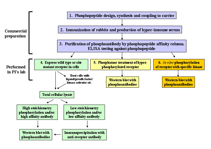
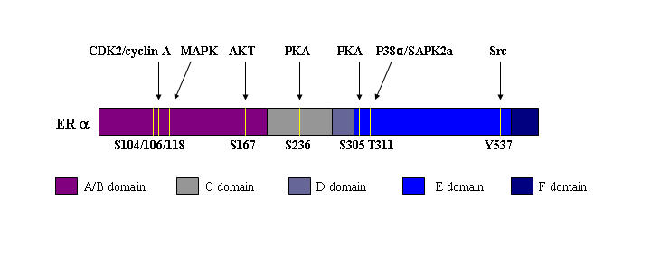
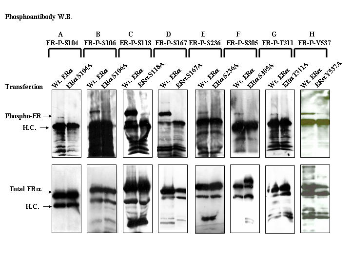
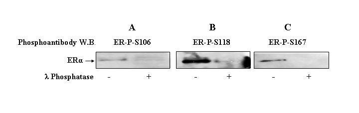
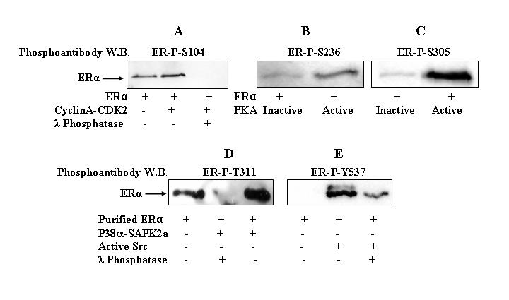
An understanding of posttranslational events in nuclear receptor signaling is crucial for drug design and clinical therapeutic strategies. Phosphorylation is a well-characterized posttranslational modification that regulates subcellular localization and function of nuclear receptors and coregulators. Although the role of single phosphorylation sites in nuclear receptor function has been described, the contribution of combinations of multiple phosphorylation sites to receptor function remains unclear. The development of phosphoantibodies to each phosphorylation site in a nuclear receptor is a powerful tool to address the role of phosphorylation in multiply phosphorylated receptors. However, phosphoantibodies must be rigorously validated prior to use. This review describes the general methodology for design, characterization and validation of phosphoantibodies using the example of eight phosphoantibodies raised against phosphorylation sites in estrogen receptor alpha (ERalpha).


Figures

Figure 1
Figure 1. Steps in phosphoantibody production and validation.
Phosphopeptide design, rabbit immunization, phosphoantibody production and affinity purification (steps 1-3) are performed by commercial vendors. Phosphoantibody specificity experiments (steps 4-6) are performed by the investigator.
Figure 2
Figure 2. Schematic representation of ERα phosphorylation sites.
ERα phosphorylation sites that have been confirmed in previous studies by in vivo and/or in vitro phosphorylation are shown.
Figure 3
Figure 3. Immunoprecipitation of wild type and mutant ERα and Western blotting with phosphoantibodies.
COS-1 cells were plated in DMEM media containing 5% FBS treated with charcoal coated dextran to remove the endogenous steroids. Cells were transfected with wild type ERα (A-H) or ERα phosphorylation site mutants ERαS104A (A), ERαS106A (B), ERαS118A (C), ERαS167A (D), ERαS236A (E), ERαS305A (F), ERαT311A (G), or ERαY537A (H). Cells were incubated with 17β-estradiol (A-D, G, H) or F/I (E, F) for 2 hrs, followed by preparation of cell extracts for immunoprecipitation. Total ERα was immunoprecipitated with ERα antibody (D12, Santa Cruz) and phosphorylation of ERα was detected by Western blot using site specific phosphoantibodies. The immunoblot was stripped and re-probed by Western blot with antibody to total ERα (Clone 6F11, Vector Laboratories).
Figure 4
Figure 4. Phosphatase treatment of purified ERα and Western blotting with phosphoantibodies.
Purified ERα (200ng) was incubated in the presence or absence of phosphatase λ (200ng) at 30 °C for 1 hr and phosphorylation of ERα was detected by Western blotting using ERα-P-S106 (A), ERα-P-S118 (B), or ERα-P-S167 (C) phosphoantibodies.
Figure 5
Figure 5. in vitro phosphorylation of purified ERα and Western blotting with phosphoantibodies.
Purified ERα (200 ng) was incubated with or without CDK2-cyclin A complex (100 ng) (A), PKA (100 ng) (B, C), P38α/SAPK2a complex (100 ng) (D), or Src (100 ng) (E) in the presence or absence of λ phosphatase (200 ng) (A, D, E) for 1 hr at 30°C. PKA was inactivated by incubating the kinase at 100°C for 2 min. ERα phosphorylation was detected by Western blot analysis using ERα-P-S104 (A), ERα-P-S236 (B), ERα-P-S305 (C), ERα-P-T311 (D), or ERα-P-Y537 (E) phosphoantibodies.

References

    1. Arnold S. F., Notides A. C. An antiestrogen: a phosphotyrosyl peptide that blocks dimerization of the human estrogen receptor. Proc Natl Acad Sci U S A. 1995a;92:7475–9. - PMC - PubMed
    1. Arnold S. F., Obourn J. D., Yudt M. R., Carter T. H., Notides A. C. in vivo and in vitro phosphorylation of the human estrogen receptor. J Steroid Biochem Mol Biol. 1995b;52:159–71. - PubMed
    1. Beck C. A., Zhang Y., Altmann M., Weigel N. L., Edwards D. P. Stoichiometry and site-specific phosphorylation of human progesterone receptor in native target cells and in the baculovirus expression system. J Biol Chem. 1996;271:19546–55. - PubMed
    1. Black B. E., Vitto M. J., Gioeli D., Spencer A., Afshar N., Conaway M. R., Weber M. J., Paschal B. M. Transient, ligand-dependent arrest of the androgen receptor in subnuclear foci alters phosphorylation and coactivator interactions. Mol Endocrinol. 2004;18:834–50. - PubMed
    1. Burns R. Immunization strategies for antibody production. Methods Mol Biol. 2005;295:1–12. - PubMed

LinkOut - more resources