. 2006 Jun;116(6):1547-60.
doi: 10.1172/JCI25397.

Intermittent pressure overload triggers hypertrophy-independent cardiac dysfunction and vascular rarefaction

Affiliations

Intermittent pressure overload triggers hypertrophy-independent cardiac dysfunction and vascular rarefaction

Cinzia Perrino et al. J Clin Invest. 2006 Jun.

Abstract

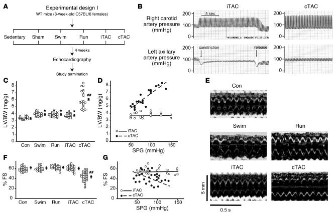
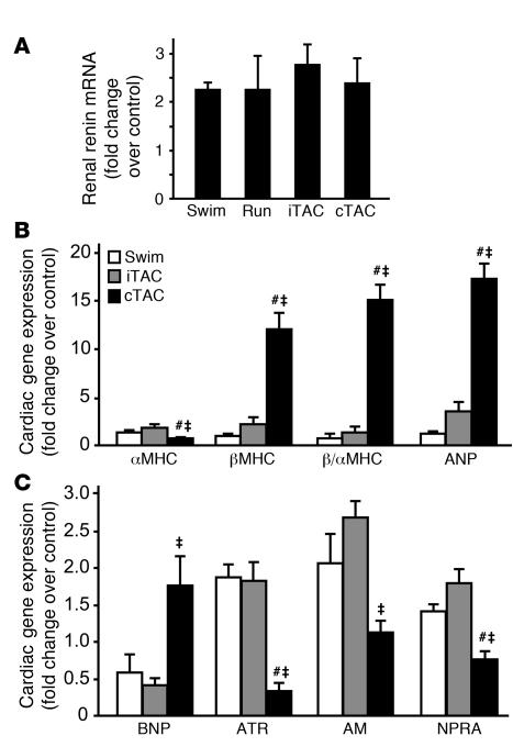
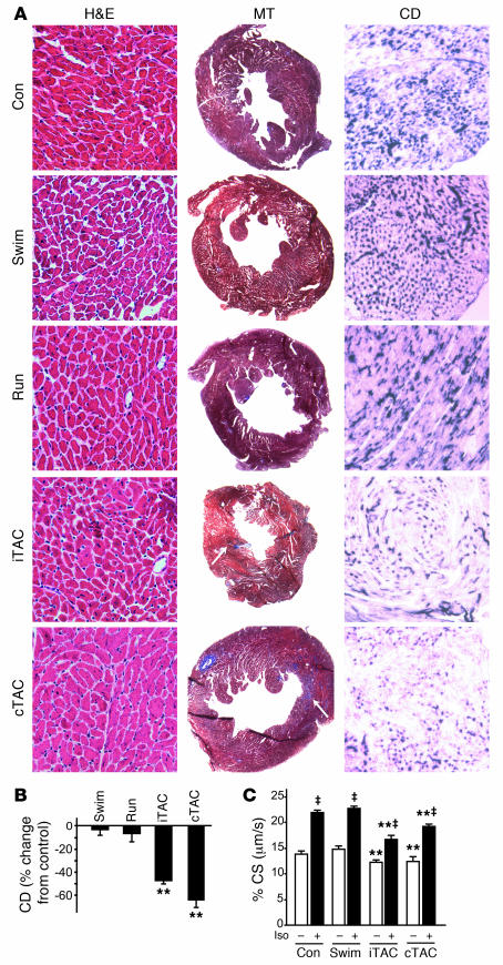
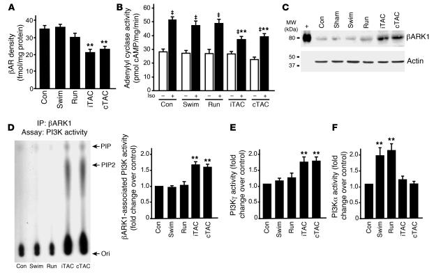
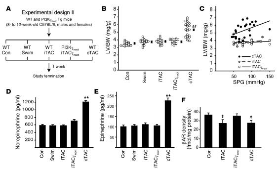
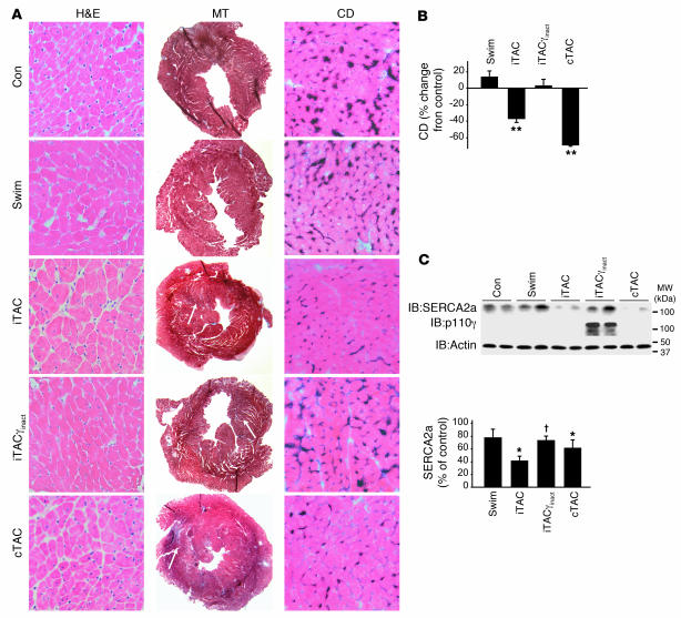
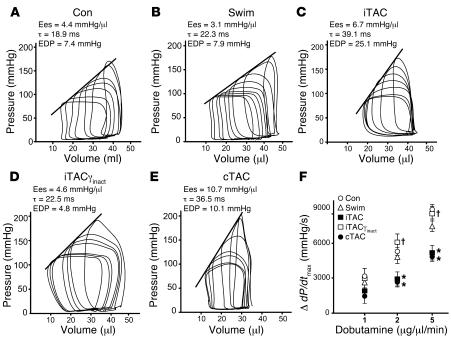
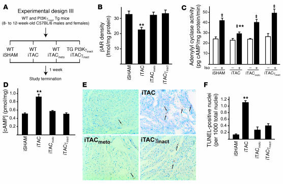
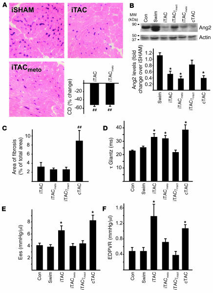
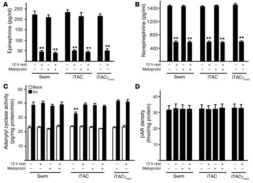
For over a century, there has been intense debate as to the reason why some cardiac stresses are pathological and others are physiological. One long-standing theory is that physiological overloads such as exercise are intermittent, while pathological overloads such as hypertension are chronic. In this study, we hypothesized that the nature of the stress on the heart, rather than its duration, is the key determinant of the maladaptive phenotype. To test this, we applied intermittent pressure overload on the hearts of mice and tested the roles of duration and nature of the stress on the development of cardiac failure. Despite a mild hypertrophic response, preserved systolic function, and a favorable fetal gene expression profile, hearts exposed to intermittent pressure overload displayed pathological features. Importantly, intermittent pressure overload caused diastolic dysfunction, altered beta-adrenergic receptor (betaAR) function, and vascular rarefaction before the development of cardiac hypertrophy, which were largely normalized by preventing the recruitment of PI3K by betaAR kinase 1 to ligand-activated receptors. Thus stress-induced activation of pathogenic signaling pathways, not the duration of stress or the hypertrophic growth per se, is the molecular trigger of cardiac dysfunction.


Figures

Figure 1
Figure 1. Duration of stress determines the magnitude of LV hypertrophy development.
(A) Experimental design of the 4-week study involving different models of physiological hypertrophy (swimming or running) or pressure overload (chronic or intermittent). Sedentary and sham-operated mice were used as controls for swimming/running and iTAC/cTAC groups, respectively, and are shown as a single control group (Con). (B) Representative tracings showing the invasive measurement of arterial pressures in the right carotid and left axillary arteries in iTAC and cTAC mice. In all iTAC animals included in the study, pulling of the externalized suture caused a rapid increase in the right carotid systolic pressure, with or without a decrease in the left axillary systolic pressure that promptly regressed after release of the suture. (C) LV/BW ratios in mice from the different groups. Filled circles with error bars indicate average ± SEM. *P < 0.05 versus control; ##P < 0.01 versus all other groups; ANOVA with Neuman-Keuls correction. (D) LV/BW ratios plotted against systolic pressure gradients measured at study termination in iTAC and cTAC mice. (E) Representative echocardiograms from the different groups of animals after 4 weeks of different protocols. (F) Percent FS in mice from the different groups. ##P < 0.01 versus all other groups; ANOVA with Bonferroni correction. (G) Percent FS plotted against systolic pressure gradients (SPG) measured at study termination in iTAC and cTAC mice.
Figure 2
Figure 2. Chronic pathological stress is required to induce the fetal gene expression reprogramming.
(A) Gene expression analysis of renal renin in kidneys of swimming, running, iTAC, and cTAC mice by RT-PCR. Data are shown as fold change over control ± SEM. Differences were not significant. (B and C) Gene expression analysis of α and β isoforms of MHC, atrial natriuretic peptide (ANP), brain natriuretic peptide (BNP), angiotensin II type 1a receptor (ATR), adrenomedullin (AM) and natriuretic peptide receptor A (NPRA) in hearts of swimming, iTAC, and cTAC mice by real-time RT-PCR of LV mRNA. Sedentary animals were used as controls for swimming mice; sham-operated animals were used for iTAC or iTAC. Data are shown as fold change over control ± SEM. #P < 0.05 versus swimming; P < 0.01 versus iTAC; Student’s t test.
Figure 3
Figure 3. Hearts exposed to physiologically applied pressure overload display a pathological structural and cellular phenotype.
(A) Representative staining of cardiac sections with H&E (magnification, ×400), MT (magnification, ×20), and endothelial alkaline phosphatase (to determine capillary density, CD; magnification, ×400). (B) Quantification of capillary density in cardiac sections stained for endothelial alkaline phosphatase expressed as percent change from control. **P < 0.01 versus control; ANOVA with Bonferroni correction. (C) Summary data for contractility studies of cardiac cells isolated from hearts of control, swimming, iTAC, and cTAC mice after 4 weeks of training (n = 5–10 mice per group; in each heart, 10–15 cells were analyzed under basal conditions and following ISO stimulation). Percent CS under basal conditions (white bars) and following ISO stimulation (black bars) is shown. P < 0.01 versus respective basal group; **P < 0.01 versus corresponding control and swimming groups; ANOVA with Neuman-Keuls correction.
Figure 4
Figure 4. βAR dysfunction is an early molecular sensor of pathological pressure overload.
(A) βAR density among membranes from hearts of control (n = 17), swimming (n = 7), running (n = 9), iTAC (n = 7), and cTAC (n = 11) mice. **P < 0.01 versus control. (B) Adenylyl cyclase activity at basal levels (white bars) and upon ISO stimulation (black bars) in hearts of control (n = 15), swimming (n = 9), running (n = 7), iTAC (n = 6), and cTAC (n = 9) mice. P < 0.01 versus respective basal group; **P < 0.01 versus control ISO; ANOVA with Bonferroni correction. (C) Myocardial lysates (80 μg) of hearts from control, swimming, running, iTAC, and cTAC mice, along with the purified protein (+), were immunoblotted for βARK1. (D) Representative PI3K assay showing βARK1-associated PI3K activity in membrane lysates from hearts of control, swimming, running, iTAC, and cTAC mice (left panel). Summary data for all the experiments performed (n = 4–7 hearts per group) are shown at right. (E and F) Summary data for PI3Kγ (E) and PI3Ka activity (F) in cytosolic lysates from the same hearts. Ori, origin; PIP, phosphatidylinositol monophosphate; PIP2, phosphatidylinositol bisphosphate. **P < 0.01 versus control; Student’s t test with Bonferroni correction.
Figure 5
Figure 5. Transgenic inhibition of βAR-targeted PI3K activity preserves βAR signaling upon intermittent pressure overload.
(A) Experimental design of the 1-week study involving WT and PI3Kγinact transgenic mice. (B) LV/BW ratios in mice from the different groups. ##P < 0.01 versus all other groups; ANOVA with Bonferroni correction. Filled circles with error bars indicate average ± SEM. (C) LV/BW ratios plotted against respective systolic pressure gradients measured at study termination in iTAC, iTACγinact, and cTAC mice. (D and E) Plasma levels of catecholamines norepinephrine (D) and epinephrine (E) in the different groups. **P < 0.01 versus control. (F) βAR density among membranes from hearts of control (n = 12), iTAC (n = 10), iTACγinact (n = 9), and cTAC (n = 11) mice. P < 0.01 versus control and iTACγinact; ANOVA with Bonferroni correction.
Figure 6
Figure 6. Preservation of βAR signaling through targeted PI3K inhibition is associated with preservation of capillary density and SERCA2a levels.
(A) Representative staining of cardiac sections with H&E (magnification, ×400), MT (magnification, ×20), and endothelial alkaline phosphatase (to determine capillary density; magnification, ×400) after 1 week of training. (B) Quantification of capillary density in cardiac sections stained for endothelial alkaline phosphatase expressed as percent reduction from control (n = 4–6 hearts per group). **P < 0.01 versus control, swimming, and iTACγinact; Student’s t test with Bonferroni correction. (C) Immunoblotting analysis of SERCA2a, actin, and PI3Kγinact protein levels in hearts from experimental design II. Densitometric quantification of SERCA2a levels is shown in the bottom panel (n = 6–10 per group). *P < 0.05 versus control, P < 0.05 versus iTAC, ANOVA with Neuman-Keuls correction.
Figure 7
Figure 7. Targeted PI3K inhibition and normalization of βAR signaling ameliorates diastolic dysfunction in hearts exposed to intermittent pressure overloads.
(AE) Representative pressure-volume loops obtained by gently pulling a suture placed around the transverse aorta in control (A), swimming (B), iTAC (C), iTACγinact (D), and cTAC (E) mice after 1 week (experimental design II). (F) Change in dP/dtmax in control (n = 11), swimming (n = 9), iTAC (n = 9), iTACγinact (n = 9), and cTAC (n = 15) groups following incremental doses (3 minutes each) of dobutamine (1, 2, and 5 μg/μl/min). *P < 0.05 versus control; Student’s t test with Bonferroni correction; P < 0.05 versus iTAC.
Figure 8
Figure 8. Beta blocker treatment preserves βAR levels and coupling in response to intermittent pressure overload.
(A) Experimental design III. (B) βAR density from the cardiac membrane of hearts from the different groups: intermittent sham-operated (iSHAM) mice, in which the iTAC suture was placed but never pulled (n = 6), as well as iTAC (n = 6), iTACmeto (n = 6), and iTACγinact mice (n = 6). (C) Adenylyl cyclase activity at basal levels (white bars) and upon ISO stimulation (black bars). P < 0.01 versus respective basal group; **P < 0.01 versus intermittent sham-operated ISO; ANOVA with Bonferroni correction. (D) Intracellular concentration of cAMP in cardiac total cell lysates. **P < 0.01 versus all groups; ANOVA with Bonferroni correction. (E) Representative TUNEL staining of cardiac sections. Arrows indicate apoptotic cell nuclei. (F) Summary data of multiple independent experiments. **P < 0.01 versus intermittent sham-operated mice.
Figure 9
Figure 9. Effects of metoprolol treatment on vascular density, fibrosis, and in vivo indices of systolic and diastolic function.
(A) Representative images of cardiac sections stained for endothelial alkaline phosphatase (magnification, ×400) and summary data of different independent experiments. (B) Representative immunoblots of cardiac lysates for angiopoietin 2 (Ang2) and relative densitometric and statistical analysis of multiple independent experiments. *P < 0.05, ##P < 0.01 versus intermittent sham-operated mice. (C) Bar graphs showing percent area of fibrosis in the different groups. ##P < 0.01 versus all other groups. (DF) Summary data for τ Glantz (D), Ees (E), and the slope of the EDPVR (F). *P < 0.05 versus control; Student’s t test with Bonferroni correction.
Figure 10
Figure 10. The combination of neurohumoral and biomechanical stress is required to trigger early βAR dysfunction in stressed hearts.
Plasma levels of catecholamines epinephrine (A) and norepinephrine (B) as well as in vitro membrane generation of cAMP (C) and βAR density (D) in cardiac membranes prepared from hearts excised immediately after termination of the training protocol or after 12 hours of rest, in the presence or absence of metoprolol before treatment. **P < 0.01 versus any stress (A and B) or versus swimming ISO and iTACγinact ISO (C).

Comment in

Similar articles

Cited by

References

    1. Meerson F.Z. Compensatory hyperfunction of the heart and cardiac insufficiency. Circ. Res. 1962;10:250–258. - PubMed
    1. Levy D., Garrison R.J., Savage D.D., Kannel W.B., Castelli W.P. Prognostic implications of echocardiographically determined left ventricular mass in the Framingham Heart Study. N. Engl. J. Med. 1990;322:1561–1566. - PubMed
    1. Frey N., Katus H.A., Olson E.N., Hill J.A. Hypertrophy of the heart: a new therapeutic target? Circulation. 2004;109:1580–1589. - PubMed
    1. Esposito G., et al. Genetic alterations that inhibit in vivo pressure-overload hypertrophy prevent cardiac dysfunction despite increased wall stress. Circulation. 2002;105:85–92. - PubMed
    1. Yusuf S., et al. Effects of an angiotensin-converting-enzyme inhibitor, ramipril, on cardiovascular events in high-risk patients. The Heart Outcomes Prevention Evaluation Study Investigators. N. Engl. J. Med. 2000;342:145–153. - PubMed

Publication types

MeSH terms

Substances