

Impact of breast density on computer-aided detection in full-field digital mammography
- PMID: 16741664
- PMCID: PMC3045151
- DOI: 10.1007/s10278-006-0592-x
Impact of breast density on computer-aided detection in full-field digital mammography
Abstract
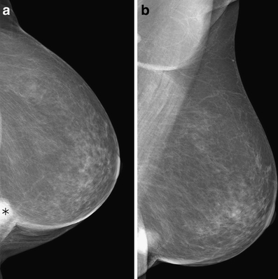
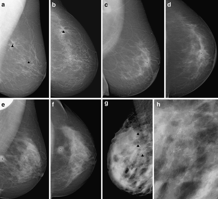
The goal of this study was to evaluate the performance of a computer-aided detection (CAD) system in full-field digital mammography (Senographe 2000D, General Electric, Buc, France) in finding out carcinomas depending on the parenchymal density. A total of 226 mediolateral oblique (MLO) and 186 craniocaudal (CC) mammographic views of histologically proven cancers were retrospectively evaluated with a digital CAD system (ImageChecker V2.3 R2 Technology, Los Altos, CA, USA). Malignant tumors were detected correctly by CAD in MLO view in 84.85% in breasts with parenchymal tissue density of the American College of Radiology (ACR) type 1, in 70.33% of the ACR type 2, in 68.12% of the ACR type 3, and in 69.70% of the ACR type 4. For the CC view, similar results were found according to the ACR types. Using the chi-square and McNemar tests, there was no statistical significance. However, a trend of better detection could be seen with decreasing ACR type. In conclusion, there seems to be a tendency for breast tissue density to affect the detection rate of breast cancer when using the CAD system.
Figures
References
-
- Moskowitz M. Screening for breast cancer: how effective are our test? A critical review. CA Cancer J Clin. 1983;33:26–39. - PubMed
-
- Bird RE. Professional quality assurance for mammography screening programs. Radiology. 1990;77:8–10. - PubMed
-
- Chan HP, Doi K, Vyborny CJ, Schmidt RA, Metz CE, Lam KL, Ogura T, Wu YZ, MacMahon H. Improvement in radiologists' detection of clustered microcalcifications on mammograms. The potential of computer-aided diagnosis. Invest Radiol. 1990;25:1102–1110. doi: 10.1097/00004424-199010000-00006. - DOI - PubMed
MeSH terms
LinkOut - more resources
Full Text Sources
Medical
Miscellaneous
