. 2006 Jun 1;20(11):1435-40.
doi: 10.1101/gad.1412806.

Persistent expression of PDX-1 in the pancreas causes acinar-to-ductal metaplasia through Stat3 activation

Affiliations

Persistent expression of PDX-1 in the pancreas causes acinar-to-ductal metaplasia through Stat3 activation

Takeshi Miyatsuka et al. Genes Dev. .

Abstract

The transcription factor pancreatic and duodenal homeobox factor 1 (PDX-1) is expressed in pancreatic progenitor cells. In exocrine pancreas, PDX-1 is down-regulated during late development, while re-up-regulation of PDX-1 has been reported in pancreatic cancer and pancreatitis. To determine whether sustained expression of PDX-1 could affect pancreas development, PDX-1 was constitutively expressed in all pancreatic lineages by transgenic approaches. The transgenic pancreas was markedly small with the replacement of acinar cells by duct-like structures, accompanied by activated Stat3. Genetic ablation of Stat3 in the transgenic pancreas profoundly suppressed the metaplastic phenotype. These results provide a mechanism of pancreatic metaplasia by which persistent PDX-1 expression cell-autonomously induces acinar-to-ductal transition through Stat3 activation.


Figures
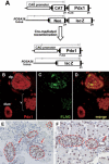
Figure 1.
Figure 1.
Cre-mediated expression of exogenous PDX-1. (A) Schematic representation of the transgenes of CAG-CAT-PDX1 and ROSA26 reporter locus and their recombination by Cre recombinase. Before recombination, the transcription of PDX-1 and lacZ is blocked by the floxed STOP cassette. When the mice are mated with Cre-expressing mice, the floxed sequence is removed by Cre recombinase, and the CAG promoter and cell type-independent ROSA26 promoter can activate the expression of PDX-1 and β-galactosidase, respectively. (pA) Polyadenylation signal. (B–D) Coimmunofluorescence for PDX-1 (red) and the Flag-tagged peptide (green) in a Ptf1a-Cre; CAG-CAT-PDX1 mouse at E10.5. The merged image demonstrates that exogenous PDX-1 is expressed only in dorsal pancreas (dp, surrounded by a dashed line), and not in duodenum (duo). Note that the red and green spots located outside the dorsal pancreas and in the vein (v) are considered to be autofluorescent signals from erythrocytes. (E,F) Immunostaining for PDX-1 in the pancreas of a control Ptf1a-Cre (E) and Ptf1a-Cre; CAG-CAT-PDX1 (F) mouse at E18.5. In the transgenic pancreas, PDX-1 is strongly expressed in >80% of cells in acini and ducts (arrows) in addition to islet cells (surrounded by a broken line).
Figure 2.
Figure 2.
Pancreatic hypoplasia in Ptf1a-Cre; CAG-CAT-PDX1 mice. (A,B) Macroscopic phenotypes of 3-wk-old pancreata of Ptf1a-Cre; ROSA26-lacZ mice (A) and the trigenic Ptf1a-Cre; CAG-CAT-PDX1; ROSA26-lacZ mice (B) stained with X-gal. Note the overall reduction in size of the trigenic pancreas.
Figure 3.
Figure 3.
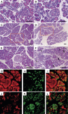
Loss of normal acinar character in Ptf1aCre; CAG-CAT-PDX1 mice. Hematoxylin and eosin (HE) staining of control and transgenic pancreata. There is no apparent difference in the morphology of 1-wk-old pancreata between Ptf1a-Cre (A) and Ptf1a-Cre; CAG-CAT-PDX1 (B) mice. Bar, 50 μm. (C–F) HE-stained sections of 3-wk-old pancreata of Ptf1a-Cre (C,E) and Ptf1a-Cre; CAG-CAT-PDX1 (D,F) mice. The boxed areas in C and D are shown at higher magnifications in E and F, respectively. (F) Abnormally shaped cells with duct-like morphology (arrows) and small acinar-like cells (arrowheads) are spread throughout the acinar area of Ptf1a-Cre; CAG-CAT-PDX1 mice. Bar, 100 μm. (G–I) Double immunostaining for PDX-1 (green) and carboxypeptidase A (red). Small PDX-1 positive cells were positive for carboxypeptidase A. (J–L) Double immunostaining for PDX-1 (green) and cytokeratin (red). PDX-1-positive duct-like cells were positive for cytokeratin.
Figure 4.
Figure 4.
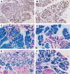
Exogenous PDX-1 expression induces acinar-to-ductal transition in a cell-autonomous manner. (A,B) Immunostaining for the Flag tag in the pancreas of a 3-wk-old Ptf1a-Cre; CAG-CAT-PDX mouse. Flag-positive cells (brown) were severely atrophic (A) or had abnormal duct-like morphology (B). X-gal staining of pancreatic sections of 1-wk-old (C,D) and 6-wk-old (E,F) mutant mice. Blue staining for lacZ expression marks cells that have undergone Cre-mediated recombination. In the pancreas of the Ptf1a-Cre; ROSA26-lacZ mouse (C), Cre-mediated recombination was observed in all acinar cells and the majority of the cells in the ductal epithelium (arrows) and the islet (surrounded by a dashed line), whereas in the pancreas of the Elastase-Cre; ROSA26-lacZ mouse (D), recombination was observed predominantly in the acinar cells, and not in the interlobular ducts (arrows) or in the islet (surrounded by a dashed line), although some terminal ductal cells (arrowheads) and a small population of islet cells were marked. Similar to the pancreas of Ptf1a-Cre; CAG-CAT-PDX1; ROSA-lacZ mice (E), there were a lot of lineage-labeled duct-like cells in the pancreas of Elastase-Cre; CAG-CAT-PDX1; ROSA26-lacZ mice (F).
Figure 5.
Figure 5.
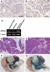
Stat3 activation is essential for acinar-to-ductal transition. (A,B) Immunostaining for tyrosine-phosphorylated Stat3 in the pancreas of a 3-wk-old Ptf1a-Cre; CAG-CAT-PDX1 mouse. Whereas none of the acinar and duct cells were positive for phosphorylated Stat3 in the control pancreas (A), a number of metaplastic cells were positive for phosphorylated Stat3 (brown) in the pancreas of Ptf1a-Cre; CAG-CAT-PDX1 mice (B). (C) Total RNA was isolated from the pancreas of CAG-CAT-PDX1 (left lane), Ptf1a-Cre (middle lane), and Ptf1a-Cre; CAG-CAT-PDX1 (right lane) mice, and subjected to RT–PCR with SOCS3 and β-actin primers. (D,E) HE-stained sections of 3-wk-old pancreata of Ptf1a-Cre; CAG-CAT-PDX1; Stat3flox/+ (D) and Ptf1a-Cre; CAG-CAT-PDX1; Stat3flox/flox mice (E). The duct-like cells seen in D are not observed in E. (F,G) Macroscopic phenotypes of 3-wk-old pancreata of Ptf1a-Cre; CAG-CAT-PDX1; Stat3flox/+; ROSA26-lacZ (F) and Ptf1a-Cre; CAG-CAT-PDX1; Stat3flox/flox; ROSA26-lacZ (G) mice. The pancreatic hypoplasia seen in F is dramatically restored in G.

References

    1. Bockman D.E. Morphology of the exocrine pancreas related to pancreatitis. Microsc. Res. Tech. 1997;37:509–519. - PubMed
    1. Edlund H. Pancreatic organogenesis—Developmental mechanisms and implications for therapy. Nat. Rev. Genet. 2002;3:524–532. - PubMed
    1. Fujitani Y., Fujitani S., Boyer D.F., Gannon M., Kawaguchi Y., Ray M., Shiota M., Stein R.W., Magnuson M.A., Wright C.V. Targeted deletion of a cis-regulatory region reveals differential gene dosage requirements for Pdx1 in foregut organ differentiation and pancreas formation. Genes & Dev. 2006;20:253–266. - PMC - PubMed
    1. Greten F.R., Weber C.K., Greten T.F., Schneider G., Wagner M., Adler G., Schmid R.M. Stat3 and NF-κB activation prevents apoptosis in pancreatic carcinogenesis. Gastroenterology. 2002;123:2052–2063. - PubMed
    1. Grippo P.J., Nowlin P.S., Cassaday R.D., Sandgren E.P. Cell-specific transgene expression from a widely transcribed promoter using Cre/lox in mice. Genesis. 2002;32:277–286. - PubMed

Publication types

Substances