. 2006 Jun 1;20(11):1441-6.
doi: 10.1101/gad.378706.

Combined ectopic expression of Pdx1 and Ptf1a/p48 results in the stable conversion of posterior endoderm into endocrine and exocrine pancreatic tissue

Affiliations

Combined ectopic expression of Pdx1 and Ptf1a/p48 results in the stable conversion of posterior endoderm into endocrine and exocrine pancreatic tissue

Solomon Afelik et al. Genes Dev. .

Abstract

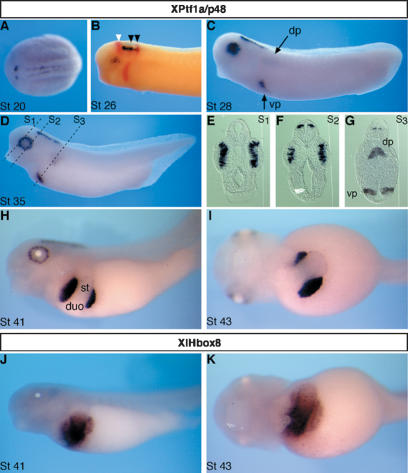
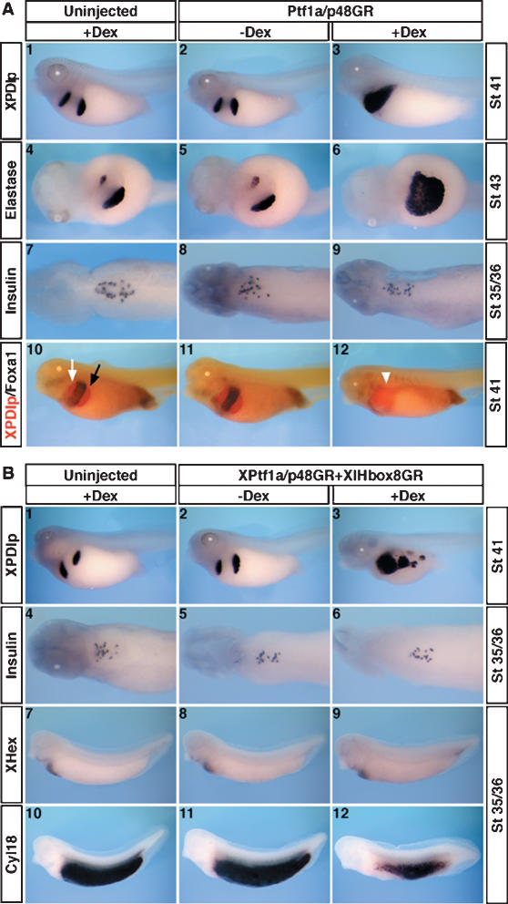
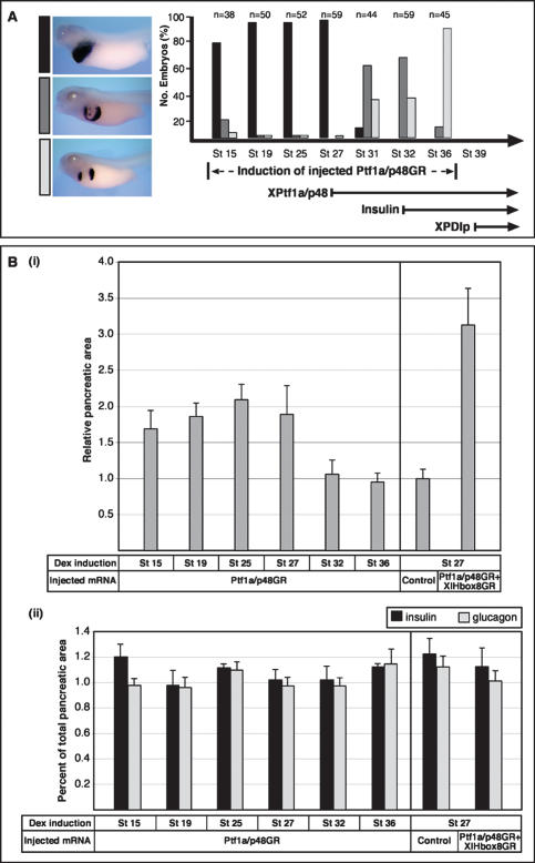
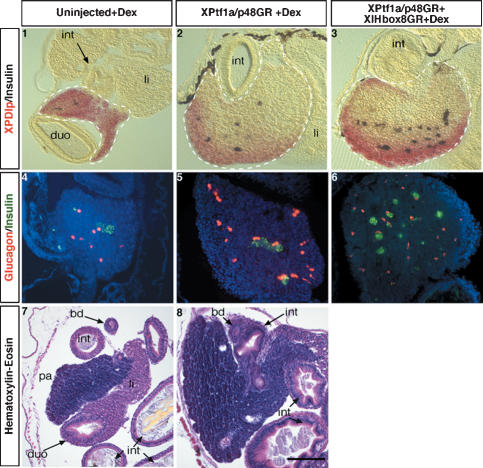
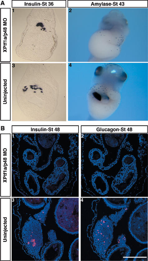
Patterning of the embryonic endoderm into distinct sets of precursor cells involves the precisely regulated activities of key transcription regulators. Ectopic, pan-endodermal activation of XPtf1a/p48 during pancreas precursor cell stages of Xenopus embryogenesis results in an expansion of the pancreatic territory, precisely within the borders of XlHbox8 expression. A combination of both activities is sufficient to expand the pancreatic precursor cell population also into more posterior portions of the endoderm. Both treatments result in the formation of a giant pancreas that persists up to late tadpole stages of development and carries both supernumerary endocrine and exocrine cells. A combination of XPtf1a/p48 and XlHbox8 is thus sufficient to convert nonpancreatic endodermal cells into pancreatic precursor cells.


Figures

Figure 1.
Figure 1.
Whole-mount in situ hybridization analysis of XPtf1a/p48 expression during Xenopus embryogenesis and in comparison to XlHbox8. (A) Dorsal view of a stage 20 embryo, anterior toward the left. XPtf1a/p48 transcripts (blue) are detected along two parallel longitudinal stripes representing the neural folds. (B) Double-staining in situ hybridization of tailbud-stage (stage 26) embryos using En2 (red) as a midbrain–hindbrain boundary marker (white arrowhead) and Krox20 (red) as a marker for rhombomeres 3 and 5 (black arrowheads). Neural expression of XPtf1a/p48 becomes restricted to the hindbrain, with the anterior end defined by the midbrain–hindbrain boundary and the posterior limit by rhombomere 5. XPtf1a/p48 transcripts are also becoming detectable in the developing retina. During later phases of development, retinal expression is confined to the proliferating precursor cells of the ciliary marginal zone and expression in the neural tube to dorsal elements (shown in E,F). (C,D) Lateral view of stage 28 and stage 35 embryos stained for XPtf1a/p48 expression. (E–G) Transverse sections (S1, S2, S3) of a stage 35 embryo at the levels indicated in D, dorsal to the top. (H) Lateral view. (I) Ventral view. (J,K) Lateral and ventral view of XlHbox8 expression. (duo) Duodenum; (dp) dorsal pancreatic bud; (st) stomach; (vp) ventral pancreatic buds.
Figure 2.
Figure 2.
A combination of XPtf1a/p48 and XlHbox8 induces ectopic pancreatic differentiation. (A) Whole-mount in situ hybridization analysis of a panel of markers reveals that ectopic expression of XPtf1a/p48 converts part of the presumptive stomach and duodenum into a pancreatic fate. (Panels 13) Lateral view. Ninety-three percent of the embryos examined showed effects as in panel 3 (n = 62). (Panels 46) Ventral view. Phenotype in panel 6, 87%, n = 24. (Panels 79) Dorsal view (head toward the left) after removal of somites, neural tube, and notochord. As shown in panel 9, none of the embryos examined exhibited altered insulin expression (n = 25). (Panels 1012) Lateral view. Double-staining in situ hybridization with Foxa1 (blue) and XPDIp (red). (Panel 10) Black and white arrows indicate dorsal and ventral pancreas, respectively. (Panel 12) The white arrowhead indicates ectopic XPDIp and loss of Foxa1 expression (85%, n = 67). (B) Combined overexpression of XPtf1a/p48 and XlHbox8 leads to a reduction of intestinal marker gene expression and concomitant ectopic expression of exocrine pancreatic marker genes in the presumptive intestine. (Panels 13,712) Lateral view. Phenotype in panel 3, 82%, n = 45; phenotype in panel 12, 70%, n = 53. (Panels 46) Dorsal view (head toward the left) after removal of somites, neural tube, and notochord.
Figure 3.
Figure 3.
The Ptf1a/p48-mediated increase of ectopic exocrine and late endocrine cells requires uncommitted endoderm. Dexamethasone induction of injected embryos was performed at different time points in between stage 15 and stage 36, as indicated. The effects on pancreas development were evaluated by whole-mount in situ hybridization analysis of XPDIp expression at stage 41 for the exocrine pancreas, and double-staining immunohistochemical analysis of insulin and glucagon expression at stage 48 for the endocrine pancreas. For morphometric quantification, the pixel number of insulin-positive and glucagon-positive cells was measured separately in serial sections using Adobe. (A) The extent of ectopic XPDIp expression at stage 41 is dependent on the stage of induction of injected Ptf1a/p48GR. The bottom right part illustrates the temporal expression profile of endogenous XPtf1a/p48, insulin, and XPDIp. (B) Despite an increase in total pancreatic area, there is no significant difference in the ratio of endocrine to exocrine pancreatic cells in embryos overexpressing Ptf1a/p48 alone or in combination with XlHbox8. (Panel i) Ectopic expression of Ptf1a/p48GR alone or in combination with XlHbox8GR results in a roughly two- and threefold increase in total pancreatic area relative to uninjected control embryos, respectively. (Panel ii) No significant difference is observed for the ratio of endocrine to total pancreatic area in a comparison of different time points for dexamethasone treatment. Sectioned pancreatic tissue was immunostained for insulin and glucagon, nuclei were counterstained with DAPI. Boundaries of the pancreatic area were delineated on the basis of morphology and pixel quantification was performed using Adobe Photoshop. An entire series of sections was analyzed for eight different embryos in each experiment. The average total pixel number of a series of pancreatic sections from control embryos is referred to as 1.
Figure 4.
Figure 4.
Giant pancreata containing both differentiated endocrine and exocrine cells at late tadpole stage are generated by ectopic expression of XPtf1a/p48GR alone or in combination with XlHbox8GR. (Panels 13) Vibratome sections of stage 48 embryos (microinjected as indicated and dexamethasone-treated at stage 27) after double-staining whole-mount in situ hybridization (insulin in blue, XPDIp in red). Due to incomplete probe penetration into the giant pancreas, the inner part remains refractory to the staining procedure; the boundaries of pancreatic tissue (broken line) were identified by microscopic analysis. Phenotype in panel 2, 60%, n = 15; in panel 3, 66%, n = 12. (Panels 46) Double-staining, immunohistochemical analysis for insulin (green) and glucagon (red) expression. In panel 5, 70%, n = 10; in panel 6, 62%, n = 8 (morphometric quantification, see Fig. 3). Bar, 200 μm. (Panels 7,8) Histological analysis (hematoxylin and eosin staining) of pancreas and neighboring tissues. In panel 8, 58%, n = 12. (bd) Bile duct; (duo) duodenum; (int) intestine; (li) liver; (pa) pancreas.
Figure 5.
Figure 5.
Early insulin expression is independent of XPtf1a/p48, while later pancreatic insulin and glucagon expression depends on XPtf1a/p48. (A) Knockdown of XPtf1a/p48 by antisense MO injection results in a loss of exocrine pancreatic gene expression, while early dorsal insulin expression remains unaffected. (Panels 1,3) Transverse vibratome sections (panel 1, 100%, n = 85; morphometric quantification of insulin expression, average pixel number in panel 1, 12,265 ± 5152; panel 3, 15,537 ± 2108). (Panels 2,4) Ventral view, head toward the top (panel 2, 77%, n = 27). (B) The second phase of endocrine pancreatic differentiation is blocked upon knockdown of XPtf1a/p48 by antisense MO injection. Immunohistochemical analysis of insulin and glucagon expression in uninjected control (panels 3,4) and in XPtf1a/p48 MO-injected embryos (panels 1,2) at stage 48 of development. Phenotype in panels 1 and 2, 60%, n = 15. Bar, 200 μm.

References

    1. Afelik S., Chen Y., Pieler T. Pancreatic protein disulfide isomerase (XPDIp) is an early marker for the exocrine lineage of the developing pancreas in Xenopus laevis embryos. Gene Expr. Patterns. 2004;4:71–76. - PubMed
    1. Ahlgren U., Jonsson J., Edlund H. The morphogenesis of the pancreatic mesenchyme is uncoupled from that of the pancreatic epithelium in IPF1/PDX1-deficient mice. Development. 1996;122:1409–1416. - PubMed
    1. Ber I., Shternhall K., Perl S., Ohanuna Z., Goldberg I., Barshack I., Benvenisti-Zarum L., Meivar-Levy I., Ferber S. Functional, persistent, and extended liver to pancreas transdifferentiation. J. Biol. Chem. 2003;278:31950–31957. - PubMed
    1. Beres T.M., Masui T., Swift G.H., Shi L., Henke R.M., MacDonald R.J. PTF1 is an organ-specific and Notch-independent basic helix–loop–helix complex containing the mammalian Suppressor of Hairless (RBP-J) or its paralogue, RBP-L. Mol. Cell. Biol. 2006;26:117–130. - PMC - PubMed
    1. Bort R., Zaret K. Paths to the pancreas. Nat. Genet. 2002;30:85–86. - PubMed

Publication types

LinkOut - more resources