. 2006 Jun 1;20(11):1525-38.
doi: 10.1101/gad.368706.

Genetic and pharmacological evidence that a retinoic acid cannot be the RXR-activating ligand in mouse epidermis keratinocytes

Affiliations

Genetic and pharmacological evidence that a retinoic acid cannot be the RXR-activating ligand in mouse epidermis keratinocytes

Cécile Calléja et al. Genes Dev. .

Abstract

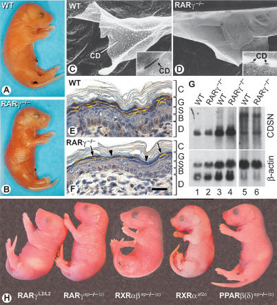
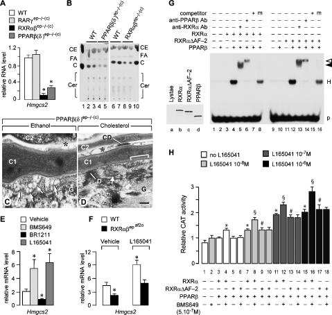
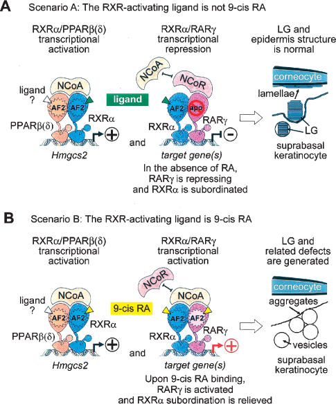
Using genetic and pharmacological approaches, we demonstrate that both RARgamma/RXRalpha heterodimers involved in repression events, as well as PPARbeta(delta)/RXRalpha heterodimers involved in activation events, are cell-autonomously required in suprabasal keratinocytes for the generation of lamellar granules (LG), the organelles instrumental to the formation of the skin permeability barrier. In activating PPARbeta(delta)/RXRalpha heterodimers, RXRalpha is transcriptionally active as its AF-2 activation function is required and can be inhibited by an RXR-selective antagonist. Within repressing RARgamma/RXRalpha heterodimers, induction of the transcriptional activity of RXRalpha is subordinated to the addition of an agonistic ligand for RARgamma. Thus, the ligand that possibly binds and activates RXRalpha heterodimerized with PPARbeta(delta) cannot be a retinoic acid, as it would also bind RARgamma and relieve the RARgamma-mediated repression, thereby yielding abnormal LGs. Our data also demonstrate for the first time that subordination of RXR transcriptional activity to that of its RAR partner plays a crucial role in vivo, because it allows RXRs to act concomitantly, within the same cell, as heterodimerization partners for repression, as well as for activation events in which they are transcriptionally active.


Figures

Figure 1.
Figure 1.
(A,B) Living wild-type (WT) and RARγ−/− newborns. Note the dull and glossy appearance of wild-type and RARγ−/− newborns, respectively. (C,D) Corneodesmosome (CD) alterations in RARγ−/− newborns. STEM views of squames. Insets show TEM views of CD. (E,F) Corneodesmosin (CDSN) distribution revealed by IHC (yellow, false color). In RARγ−/−, some granular keratinocytes contain CDSN (arrows), whereas others display less or no CDSN (arrowheads). The section in E is slightly tangential when compared with F, resulting in an oval appearance of nuclei. (G) Northern blot of total RNA (25 μg) from esophagus (lanes 1,2), tongue (lanes 3,4), and back skin (lanes 5,6). Note that two CDSN RNA species are detected in skin. The blot was also hybridized with a β-actin probe. (H) Newborn external appearance. The skin surface appears dull in controls (RARγL2/L2), RXRαaf2o, and PPARβ(δ)ep−/−(c) mutants, but glossy in RARγep−/−(c) and RXRαβep−/−(c) mutants. (B) Basal layer; (C) cornified layer; (CD) corneodesmosome; (D) dermis; (G) granular layer; (S) spinous layer. Bar in F represents 15 μm in C and D and 50 μm in E and F.
Figure 2.
Figure 2.
(A–I) Ultrastructure of granular keratinocytes in wild-type (WT) and RARγ−/− newborns. (A,B) In wild type, numerous LGs (large arrows) with conspicuous internal lamellar structures (inset) are present, while vesicles that lack (inset, asterisk) or display disorganized lamellae (thin arrows) are observed in RARγ−/−. (C–E) Detection of acid lipase activity at birth. The arrowhead in C points to a LG containing the electron-dense “dotted” lipase reaction product. Incubation without substrate was used as a negative control in E; the large arrow points to a LG. (F,G) Junction between the outermost granular and the first cornified layer. Large arrows point to wild-type LGs that fuse with the granular keratinocyte membrane to release their lamellar content at the cell surface (bracket). Thin arrows point to vesicle aggregates in RARγ−/−. (H,I) Large CDs connect wild-type corneocytes together, and lipid-rich multilamellar sheets fill the space between corneocytes (inset). Smaller CDs and aggregates of vesicles are present between the corneocytes (thin arrows) in RARγ−/−. (J–R) Ultrastructure of granular keratinocytes in adult mice bearing somatic mutations. (J) In Tam-treated control mice, granular keratinocytes contain LGs that release their lipid lamellar content at the cell surface (large arrows), and large CDs connect the corneocytes together. (K,L) In RARγep−/− and RARγsb−/− mice, vesicles form aggregates at the apical pole of granular keratinocytes (thin arrows), while CDs are small. (M–R) Similar alterations are present in RXRαep−/−(c), RXRαβep−/−(c), RXRαβepaf2o(c), and PPARβ(δ)sb−/− mice, whereas in PPARα−/− and PPARγep−/−(c) mice, granular keratinocytes contain normal LGs releasing their lamellar content at the cell surface (large arrows). Basal and spinous keratinocytes always appeared normal (not shown). C1, C2, and C3 indicate the first, second, and third cornified layers, respectively. (CD) Corneodesmosome; (D) desmosome; (G) granular keratinocyte; (KF) keratin filament bundle; (KG) keratohyalin granule; (M) mitochondria. Bar in O represents 0.5 μm in A and B; 0.1 μm in the insets in A, B, and C–E; and 0.15 μm in F–R.
Figure 3.
Figure 3.
Histochemical detection of lipids in newborn (A,B) and adult (C–N) mice, using Nile red staining. (A–H) On top of the cornified layer, neutral lipids are concentrated as a yellow continuous ribbon in wild-type (WT) epidermis, while their distribution is interrupted (arrowheads) in mutants as indicated. The dermis staining in C is artifactual. (I–N) Surface lipid distribution is interrupted (arrowheads) in wild-type mice topically treated with RA, BMS961, or BR1211, whereas it remains continuous upon topical treatment with BMS753, BMS649, or BMS493. Note in I the typical thickening of the epidermis resulting from RA-induced basal keratinocyte proliferation (Chapellier et al. 2002b). Phospholipid distribution (orange) is not affected. (B) Basal layer; (C) cornified layer; (D) dermis; (E) epidermis; (SB) suprabasal layer. Bar in N represents, 50 μm in A and B and 300 μm in C–N.
Figure 4.
Figure 4.
Epidermis ultrastructure in adult mice topically treated with retinoids. (A–R) TEM views from mice treated as indicated using acetone (vehicle control), RA, BMS753, BMS961, BMS649 alone or in combination with BMS961, BMS493, and BR1211 alone or in combination with BMS649. In control, BMS753-, BMS649-, and BMS493-treated mice, normal LGs releasing their lipid content at granular keratinocyte apical pole (large arrows) to form multilamellar sheets between corneocytes (brackets) are observed. In RA-, BMS961-, and BR1211-topically treated mice, vesicles (thin arrows) form aggregates at the granular keratinocyte apical pole, and persist between corneocytes (asterisks). Coadministration of BMS649 and BMS961 worsens the defects. Coadministration of BMS649 mostly prevents the appearance of the BR1211-induced defects, although few aggregates can be observed between corneocytes (asterisk in O). C1 and C2 indicate the first and second cornified layers, respectively. (D) Desmosome; (G) granular keratinocyte. Bar in R represents 0.1 μm in A–C, G–I, and M–O, and 0.5 μm in D–F, J–L and P–R.
Figure 5.
Figure 5.
(A–F) Alteration of cholesterol metabolism upon PPARβ(δ) or RXRα (and β) ablations in keratinocytes. (A) Real-time quantitative RT–PCR analysis for Hmgcs2 transcripts in total RNA from wild-type (WT), RARγep−/−(c), RXRαβep−/−(c), and PPARβ(δ)ep−/−(c) newborn epidermis. Values (arbitrary units) correspond to the mean amount ± SEM of RNA transcripts detected in the epidermis of each genotype (n = 4), relative to the amount of β-actin transcripts (unchanged upon mutation). (B) TLC analysis of lipids from the surface of newborns with genotypes as indicated. (Lanes 1,10) Lipid standards. (C) Cholesterol; (CE) cholesteryl ester (stearate); (Cer) ceramides; (FA) fatty acids. (C,D) TEM views from PPARβ(δ)ep−/−(c) epidermis topically treated with cholesterol. (C) In ethanol (vehicle)-treated mutants, vesicles aggregate at the granular keratinocyte apical pole and persist between corneocytes (asterisk). (D) In cholesterol-treated mutants, LGs release their lipid content at the granular keratinocytes apical pole to form multilamellar sheets (brackets). Aggregates of vesicles present between corneocytes (asterisk) arise from the PPARβ(δ)-null keratinocytes that differentiated into corneocytes before the onset of cholesterol administration. C1 and C2 indicate the first and second cornified layers, respectively. (CD) Corneodesmosome; (D) desmosome; (G) granular keratinocyte. Bar in D represents 0.1 μm. (E) Real-time quantitative RT–PCR analysis for Hmgcs2 transcripts in total RNA from back skin of wild-type adult mice topically treated with acetone vehicle, BMS649, BR1211, or L165041. Values (arbitrary units) correspond to the mean amount ± SEM of RNA transcripts detected in each series (n = 3), relative to the amount of glyceraldehyde-3-phosphate dehydrogenase transcripts (unchanged upon topical treatment). (F) Real-time quantitative RT–PCR analysis for Hmgcs2 transcripts in total RNA from the back skin of wild-type and RXRαβepaf2o mice treated with ethanol vehicle or L165041. Values (arbitrary units) correspond to the mean amount ± SEM of RNA transcripts detected in the skin of each genotype (n = 3), relative to the amount of 36B4 transcripts (unchanged upon mutation). (A,E,F) Asterisks indicate a significant difference from the wild-type values (p < 0.05). (G,H) Effects of AF-2 deletion on binding property and transcriptional activity of RXRα in heterodimer with PPARβ(δ). (G) EMSA showing that both RXRα/PPARβ(δ) (lane 4) and RXRαΔAF-2/PPARβ(δ) (lane 12) heterodimers (H) equally bind to the Hmgcs2 PPRE (p). The complexes formed were supershifted by addition of antibodies against RXRα (closed arrowhead, lanes 5,13) or PPARβ(δ) (open arrowhead, lanes 6,14). EMSA competitions were performed with a 1000-fold excess of unlabeled PPRE (lanes 7,15) or mutated PPRE (m, lanes 8,16). The left inset shows SDS-PAGE analysis of in vitro translated proteins, indicating that RXRα (lane b), RXRαΔAF-2 (lane c), and PPARβ(δ) (lane d) were produced with similar efficiencies. Lane a contains the same amount of lysate translated without receptor cDNA template. (H) HepG2 cell transfections with the Hmgcs2 PPRE-tk-Cat reporter, and RXRα-, RXRαΔAF-2-, and PPARβ(δ)-expressing vectors, as indicated. The ligand for RXRα (BMS649) was added at 5.10−7 M, as indicated. The ligand for PPARβ(δ) (L165041) was added at increasing concentrations: 10−8 M (gray bars), 10−7 M (dark-gray bar), and 10−6 M (black bars). (White bars) No L165041 added. Four independent experiments were performed with triplicate transfections for each condition. Bars show the mean CAT activity ± SEM relative to β-galactosidase activity. (*,§,#) Indicate a significant difference versus lane 3 (p < 0.01), lane 4 (p < 0.001), and lane 5 (p < 0.01), respectively. There is no significant change between lanes 3, 5, and 6; lanes 7, 9, and 10; lanes 11, 13, and 14; and lanes 15, 17, and 18.
Figure 6.
Figure 6.
Effects of PPARβ(δ) activation on epidermis ultrastructure and surface lipid distribution in adult mice with genotypes as indicated. TEM views (A–H) and histochemical detection of lipids using Nile red staining (I–L) in control mice (RXRαβL2/L2) and in mutants expressing RXRα and RXRβ lacking their AF-2 (RXRαβepaf2o), topically treated with acetone vehicle or with the PPARβ(δ)-selective agonist L165041. (A,B,E,F,I,J) In controls, normal LGs (large arrows) release their content at the granular keratinocyte apical pole, and lipid distribution is even. (C,G,K) In RXRαβepaf2o mice treated with acetone, vesicles (thin arrows) form aggregates that persist between corneocytes, and lipid distribution is interrupted (arrowheads). (D,H,L) Topical administration of L165041 to RXRαβepaf2o mice cures LG and surface lipid defects. C1 and C2 indicate the first and second cornified layers, respectively. (CD) Corneodesmosomes; (D) desmosome; (G) granular keratinocyte; (M) mitochondria. Bar in L represents 0.1 μm in A–D, 0.5 μm in E–H, and 160 μm in I–L.
Figure 7.
Figure 7.
The concomitant occurrence of RAR/RXR-mediated repression and RXR/PPARβ(δ)-mediated activation events observed in mouse epidermis suprabasal keratinocytes refutes the possibility that the ligand activating RXR AF-2 could be 9-cis RA. (A) Scenario A: The RXR-activating ligand is not 9-cis RA. Within activating PPARβ(δ)/RXRα heterodimers, the AF-2 of RXRα is required to activate gene expression, notably that of Hmgcs2. Thus, RXRα most likely binds an agonistic ligand, and PPARβ(δ)/RXRα heterodimers interact with coactivators (NcoAs/SRCs) (shown in the left panel). On the other hand, within repressing RARγ/RXRα heterodimers, RXRα cannot be transcriptionally active, due to subordination of its transcriptional activity to that of its repressing RARγ partner, which has to be in its unliganded apo-form in order to bind corepressors (NcoR/SMRT) that block the transcriptional activity of RXRα (shown in the middle panel). It follows that there should not be any retinoic acids in suprabasal keratinocytes, and therefore that the RXR-activating ligand cannot be 9-cis RA. Under these conditions, lamellar granule (LG) formation and epidermis ultrastructure are normal (shown in the right panel). (B) Scenario B: The RXR-activating ligand is 9-cis RA. In this case, RXRα binds 9-cis RA and PPARβ(δ)/RXRα heterodimers interact with coactivators (NcoAs/SRCs) (shown in the left panel). However, 9-cis RA also binds to RARγ, thus relieving RXRα subordination and activating RARγ/RXRα heterodimers, which no longer repress gene expression (shown in the middle panel). Under these conditions, LG biogenesis is impaired (shown in the right panel). As this is not actually the case, the possibility that 9-cis RA could be the ligand activating RXR AF-2 is ruled out.

References

    1. Abu-Abed S., MacLean G., Fraulob V., Chambon P., Petkovich M., Dolle P. Differential expression of the retinoic acid-metabolizing enzymes CYP26A1 and CYP26B1 during murine organogenesis. Mech. Dev. 2002;110:173–177. - PubMed
    1. Allenby G., Bocquel M.T., Saunders M., Kazmer S., Speck J., Rosenberger M., Lovey A., Kastner P., Grippo J.F., Chambon P., et al. Retinoic acid receptors and retinoid X receptors: Interactions with endogenous retinoic acids. Proc. Natl. Acad. Sci. 1993;90:30–34. - PMC - PubMed
    1. Andersen B., Rosenfeld M.G. Intracellular receptors. New wrinkles in retinoids. Nature. 1995;374:118–119. - PubMed
    1. Arnold M.L., Anton-Lamprecht I., Melz-Rothfuss B., Hartschuh W. Ichthyosis congenita type III. Clinical and ultrastructural characteristics and distinction within the heterogeneous ichthyosis congenita group. Arch. Dermatol. Res. 1988;280:268–278. - PubMed
    1. Attar P.S., Wertz P.W., McArthur M., Imakado S., Bickenbach J.R., Roop D.R. Inhibition of retinoid signaling in transgenic mice alters lipid processing and disrupts epidermal barrier function. Mol. Endocrinol. 1997;11:792–800. - PubMed

Publication types

LinkOut - more resources