. 2006 Sep 1;91(5):1630-7.
doi: 10.1529/biophysj.106.080721. Epub 2006 Jun 2.

Molecular dynamics study of MscL interactions with a curved lipid bilayer

Affiliations

Molecular dynamics study of MscL interactions with a curved lipid bilayer

Grischa R Meyer et al. Biophys J. .

Abstract

Mechanosensitivity is a ubiquitous sensory mechanism found in living organisms. The simplest known mechanotransducing mechanism is found in bacteria in the form of the mechanosensitive membrane channel of large conductance, MscL. This channel has been studied extensively using a variety of methods at a functional and structural level. The channel is gated by membrane tension in the lipid bilayer alone. It serves as a safety valve protecting bacterial cells against hypoosmotic shock. MscL of Escherichia coli embedded in bilayers composed of asymmetric amounts of single-tailed and double-tailed lipids has been shown to gate spontaneously, even in the absence of membrane tension. To gain insight into the effect of the lipid membrane composition and geometry on MscL structure, a fully solvated, all-atom model of MscL in a stress-free curved bilayer composed of double- and single-tailed lipids was studied using a 9.5-ns molecular dynamics simulation. The bilayer was modeled as a domed structure accommodating the asymmetric composition of the monolayers. During the course of the simulation a spontaneous restructuring of the periplasmic loops occurred, leading to interactions between one of the loops and phospholipid headgroups. Previous experimental studies of the role of the loops agree with the observation that opening starts with a restructuring of the periplasmic loop, suggesting an effect of the curved bilayer. Because of limited resources, only one simulation of the large system was performed. However, the results obtained suggest that through the geometry and composition of the bilayer the protein structure can be affected even on short timescales.


Figures

FIGURE 1
FIGURE 1
(A) Two-dimensional representation of the curved bilayer. The curvature is induced by a heterogeneous distribution of single-tailed lipids (pink), that form the upper (lower) monolayer in regions I (II), and doubletailed lipids (gray), that form the lower (upper) monolayer in regions I (II). The quantities shown, r, t, ϕ, are used to describe mathematically the curved bilayer (see Methods). (B) A slice through the center of the protein-lipid-water system after 2 ns of simulation. Lipids and the MscL protein are color-coded as in C, except that the periplasmic loops are shown in orange. Water molecules are presented in turquoise. (C) Stereo view of the lipid bilayer and the MscL protein before equilibration. Water molecules are not shown. Single-tailed lipids are presented in pink, double-tailed lipids in gray. MscL is colored as in Fig. 3.
FIGURE 2
FIGURE 2
Time development of the RMSD of MscL. The RMSD values shown were determined for the Cα-atoms only and correspond to the whole protein (solid line), the periplasmic loops (dashed line), the transmembrane helices (dotted line), the gating region, i.e., residues A-20 to A-28 (dash-dash-dotted line), and the N-terminal (S1) helices (dash-dotted line).
FIGURE 3
FIGURE 3
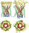
MscL at the beginning (left) and end (right) of the 9.5-ns simulation. Shown are the N-terminal helices S1 (blue), the transmembrane helices TM1 (red), the periplasmic helices S2 (orange), the periplasmic loops S2 (yellow), and the transmembrane helices TM2 (green). The C-terminus (S3) has been cut off in our simulation and is not shown.
FIGURE 4
FIGURE 4
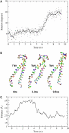
(A) Rotation of the gating region of subunit 1 (residues A-20 to A-28). The gating region is seen to rotate during the simulation relative to the remaining MscL. The dots represent snapshots from the simulation; the line represents a running average (over a 100-ps window). (B) Conformational dynamics of subunit 1. Shown are conformations of the subunit (helices TM1 and TM2) at the beginning of the simulation, at 3.3 ns, and at the end of the simulation. (C) Time development of the distance between hydrogen-bonding atoms of A-28 and I-32.
FIGURE 5
FIGURE 5
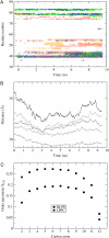
(A) Time development of lipid-protein interaction. For each event in which lipid headgroups and protein side groups approach each other to <3 Å, a symbol is shown for the respective side group. Colors differentiate side groups of subunit 1 (black circle), 2 (red square), 3 (green diamond), 4 (blue cross), and 5 (orange triangle). (B) Thickness of the lipid bilayer. The thickness is measured as the distance between the phosphorus atoms. Shown are averages encompassing the phosphorus atoms at radii 10 Å (solid), 20 Å (dotted), 30 Å (dashed), and 40 Å (dash-dotted) from the protein, and all phosphorus atoms (dash-dash-dotted). (C) Deuterium order parameter SCD = 〈3/2 cos2 θ – 1/2〉, where θ is the angle between each C-H bond and the bilayer normal. Shown are the order parameters at each acyl chain carbon for the DLPE (double-tailed) and LLPC (single-tailed) lipids.

References

    1. Martinac, B. 2004. Mechanosensitive ion channels: molecules of mechanotransduction. J. Cell Sci. 117:2449–2460. - PubMed
    1. Hamill, O., and B. Martinac. 2001. Molecular basis of mechanotransduction in living cells. Physiol. Rev. 81:685–740. - PubMed
    1. Sukharev, S., W. Sigurdson, C. Kung, and F. Sachs. 1999. Energetic and spatial parameters for gating of the bacterial large conductance mechanosensitive channel, MscL. J. Gen. Physiol. 113:525–540. - PMC - PubMed
    1. Häse, C., A. Le Dain, and B. Martinac. 1995. Purification and functional reconstitution of the recombinant large mechanosensitive ion channel (MscL) of Escherichia coli. J. Biol. Chem. 270:18329–18334. - PubMed
    1. Maurer, J., and D. Dougherty. 2003. Generation and evaluation of a large mutational library from the Escherichia coli mechanosensitive channel of large conductance, MscL: implications for channel gating and evolutionary design. J. Biol. Chem. 278:21076–21082. - PubMed

Publication types

LinkOut - more resources