. 2006 Jun 13;103(24):9292-7.
doi: 10.1073/pnas.0602135103. Epub 2006 Jun 2.

Conformational suppression of inter-receptor signaling defects

Affiliations

Conformational suppression of inter-receptor signaling defects

Peter Ames et al. Proc Natl Acad Sci U S A. .

Abstract

Motile bacteria follow gradients of attractant and repellent chemicals with high sensitivity. Their chemoreceptors are physically clustered, which may enable them to function as a cooperative array. Although native chemoreceptor molecules are typically transmembrane homodimers, they appear to associate through their cytoplasmic tips to form trimers of dimers, which may be an important architectural element in the assembly and operation of receptor clusters. The five receptors of Escherichia coli that mediate most of its chemotactic and aerotactic behaviors have identical trimer contact residues and have been shown by in vivo crosslinking methods to form mixed trimers of dimers. Mutations at the trimer contact sites of Tsr, the serine chemoreceptor, invariably abrogate Tsr function, but some of those lesions (designated Tsr*) are epistatic and block the function of heterologous chemoreceptors. We isolated and characterized mutations (designated Tar()) in the aspartate chemoreceptor that restored function to Tsr* receptors. The suppressors arose at or near the Tar trimer contact sites and acted in an allele-specific fashion on Tsr* partners. Alone, many Tar() receptors were unable to mediate chemotactic responses to aspartate, but all formed clusters with varying efficiencies. Most of those Tar() receptors were epistatic to WT Tsr, but some regained Tar function in combination with a suppressible Tsr* partner. Tar()-Tsr* suppression most likely occurs through compensatory changes in the conformation or dynamics of a mixed receptor signaling complex, presumably based on trimer-of-dimer interactions. These collaborative teams may be responsible for the high-gain signaling properties of bacterial chemoreceptors.


Conflict of interest statement

Conflict of interest statement: No conflicts declared.

Figures

Fig. 1.
Fig. 1.
Working model of inter-receptor epistasis and suppression. Tsr receptors with epistatic lesions (Tsr*) block the function of WT Tar receptors by forming defective mixed trimers of dimers. The Tsr* amino acid replacement (star) affects one of the trimer contact residues (small open circles). Tar mutations (small triangles) may impart a compensatory conformational change to mixed trimers. Tar mutations listed in italics affect trimer contact residues. Some Tar mutants retain signaling function in the presence of their Tsr* partner, whereas others do not. When tested alone, most of the nonfunctional Tar receptors are themselves epistatic (Tar*) to WT Tsr receptors.
Fig. 2.
Fig. 2.
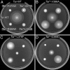
Chemotaxis phenotypes of Tsr* and Tar mutants. Strain UU1250 with various combinations of mutant Tsr and Tar plasmids was tested for chemotactic ability on tryptone soft agar. Plates were photographed after 10-h incubation at 32.5°C. (A) Epistatic behavior of Tsr*. The doubly WT control colony (center) forms two cell bands or rings reflecting serine chemotaxis (Tsr function; outer ring) and aspartate chemotaxis (Tar function; inner ring). WT Tar fails to promote aspartate taxis in cells that also carry an epistatic Tsr receptor (Tsr*). (B) Suppression by Tar-V396A. This Tar mutant functions well with WT Tsr and restores partial function to Tsr*-I377A and full function to Tsr*-R388W and L380A. Note that the three largest colonies also contain an aspartate taxis ring. (C) Epistasis and suppression by Tar-L376F. This Tar mutant is fully epistatic to WT Tsr but suppresses Tsr*-F373W and (less well) R388W. The suppressed colonies have a serine ring but lack an aspartate ring. (D) Epistasis and suppression by Tar-G388D. This Tar mutant is moderately epistatic to WT Tsr but specifically suppresses Tsr*-L380A. The suppressed colony has both a serine ring and an aspartate ring.
Fig. 3.
Fig. 3.
Trimer contact regions of Tsr and Tar receptors. Both structures depict backbone traces of the cytoplasmic tip of a receptor dimer (Tsr residues 361–420), showing residues at the trimer interface. The two subunits are identical but given different thicknesses to indicate their different structural environments in the trimer of dimers. The thicker subunit contributes most of the residue contacts at the trimer interface. Those same residues (atoms not shown) in the other subunit are arrayed on the outside of the trimer. For Tsr, the 11 space-filled residues comprise the principal trimer contact sites. Residues on the right define the major trimer contact helix. Amino acid replacements at any of the trimer contact sites can create epistatic behavior, but only the four with dark shading were suppressible by Tar receptors. For Tar, space-filled alpha carbons denote the residues at which Tar mutations were obtained. Residues on the right define the major trimer contact helix. Tar amino acid replacements with WT signal output are shaded light gray; counter-clockwise-biased mutations are shaded white; CW-biased mutations are shaded dark gray. Tar mutations listed in italics affect trimer contact residues.
Fig. 4.
Fig. 4.
Nonadditivity of Tsr* and Tar flagellar rotation patterns. In each row, the extent of CW rotational bias produced by Tsr* receptors alone is indicated by stars, and the rotational bias produced by each of their Tar suppressors alone is indicated by circles. The WT label identifies the rotational bias of cells carrying both WT Tsr and WT Tar receptors.
Fig. 5.
Fig. 5.
Examples of conformational suppression mechanisms in receptor trimers of dimers. Backbone traces of the trimer contact region of three different trimers, viewed from the cytoplasmic tip. Each trimer contains one Tsr* dimer (dark gray) and two Tar dimers (white). The black space-filled residues in Tsr indicate the trimer contact residue altered by the Tsr* mutation; white atoms indicate the corresponding (WT) residue in the Tar receptors. Light-gray space-filled atoms in Tar indicate the residue changed by the Tar alteration; dark gray atoms indicate its (WT) counterpart in the Tsr* receptor.

References

    1. Zhulin I. B. Adv. Microb. Physiol. 2001;45:157–198. - PubMed
    1. Sourjik V. Trends Microbiol. 2004;12:569–576. - PubMed
    1. Parkinson J. S., Ames P., Studdert C. A. Curr. Opin. Microbiol. 2005;8:116–121. - PubMed
    1. Block S. M., Segall J. E., Berg H. C. Cell. 1982;31:215–226. - PubMed
    1. Jasuja R., Lin Y., Trentham D. R., Khan S. Proc. Natl. Acad. Sci. USA. 1999;96:11346–11351. - PMC - PubMed

Publication types

MeSH terms

LinkOut - more resources