. 2006 Jun 13;103(24):9001-5.
doi: 10.1073/pnas.0502718103. Epub 2006 Jun 2.

Microfibrillar structure of type I collagen in situ

Affiliations

Microfibrillar structure of type I collagen in situ

Joseph P R O Orgel et al. Proc Natl Acad Sci U S A. .

Abstract

The fibrous collagens are ubiquitous in animals and form the structural basis of all mammalian connective tissues, including those of the heart, vasculature, skin, cornea, bones, and tendons. However, in comparison with what is known of their production, turnover and physiological structure, very little is understood regarding the three-dimensional arrangement of collagen molecules in naturally occurring fibrils. This knowledge may provide insight into key biological processes such as fibrillo-genesis and tissue remodeling and into diseases such as heart disease and cancer. Here we present a crystallographic determination of the collagen type I supermolecular structure, where the molecular conformation of each collagen segment found within the naturally occurring crystallographic unit cell has been defined (P1, a approximately 40.0 A, b approximately 27.0 A, c approximately 678 A, alpha approximately 89.2 degrees , beta approximately 94.6 degrees , gamma approximately 105.6 degrees ; reflections: 414, overlapping, 232, and nonoverlapping, 182; resolution, 5.16 A axial and 11.1 A equatorial). This structure shows that the molecular packing topology of the collagen molecule is such that packing neighbors are arranged to form a supertwisted (discontinuous) right-handed microfibril that interdigitates with neighboring microfibrils. This interdigitation establishes the crystallographic superlattice, which is formed of quasihexagonally packed collagen molecules. In addition, the molecular packing structure of collagen shown here provides information concerning the potential modes of action of two prominent molecules involved in human health and disease: decorin and the Matrix Metallo-Proteinase (MMP) collagenase.


Conflict of interest statement

Conflict of interest statement: No conflicts declared.

Figures

Fig. 1.
Fig. 1.
Background subtracted off-meridian diffraction pattern of rat tail tendon (Upper) and simulated diffraction pattern from model-derived intensities (Lower). Rsim (R factor between model generated and observed diffraction pattern) was determined to be 16.7%, whereas the R factor for the integrated structure factors is 9.55% (Rw being 21.73%).
Fig. 2.
Fig. 2.
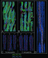
Electron density maps. Map elements in A and B have been compressed five times along the direction parallel to the c-axis for clarity. (A) Overlap region of 1D repeat, showing the common tilt of the five collagen segments within the overlap region. The triple-helical backbone of the prerefined model is shown in red as a stylized Cα trace. (B) Expanded view of the gap region showing the different paths of the gap region molecules to that of those in the overlap and to each other in the gap. Small patches of unassigned electron density are seen at 0.6D and 0.8D of the unit cell in the observed/experimental phase (Po) maps. Two large cavities are also found at these locations. The triple-helical backbone is shown in red as a stylized Cα trace, and the calculated/model phase map is shown as 2FoFc Pm (phases averaged between calculated and observed to reduce possible phase bias). (C) The electron density at the N and C telopeptide levels of the D-periodic unit cell and conformation of the telopeptides. The amino acid residue Cα trace is shown in red except for lysine/hydroxy-lysine residues, which are colored yellow. The N-telopeptide-containing segment (segment 1; Left) and C-telopeptide-containing segment (segment 5; Right) are labeled. (D) An ≈225-Å-long section from the native cell (original aspect ratio preserved, i.e., not compressed along the c-axis), showing the section of the bent collagen helix just below 0.6D of the axial unit cell. The electron density of neighboring chain segments can be seen along the side-chain traced segment. The rigid-body refined model is shown in red; the final relaxed model is in yellow.
Fig. 3.
Fig. 3.
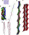
Collagen organization and structure. The collagen segments are labeled as follows for B, C, and E: 1, gray; 2, red; 3, green; 4, blue; 5, yellow. Part of segment 1 is colored cyan (the N terminus), and part of segment 5 is colored magenta (the C terminus) to allow easier identification in B. The c-axis has been compressed five times for B, C, and E. (A) Electron density and model showing the quasihexagonal packing of the molecular segments. The approximate outline of the unit cell (a and b sides) is marked with black lines. (B) Cα carbons rendered as line spheroids showing the conformation of the D-staggered collagen segments within a single unit cell (cell axis shown). (C) Molecular path of a collagen molecule through successive unit cells in the ac plane. (D) Enlarged view of the telopeptides of type I collagen, showing N-telopeptide (left and bottom of C) and C-telopeptide (right and top of C). Both have been rotated with respect to C for clarity of display. (E) Taking several 1D staggered collagen molecules from the collagen packing structure (single molecule shown in C), it is possible to represent the collagen microfibril. The collagen molecules progress from bottom to top (N to C terminus) and are colored as previously (except that chains starting in successive D-periods are darker equivalent colors). A clear right-handed twist can be seen, particularly between segments 2 and 3 (which is roughly at the midpoint of each collagen molecule). The noncrystallographic symmetry that relates the collagen molecules within the microfibrillar structure is a simple fractional translational function (Nu, 0v, Nw) where N is an integer. Five successive D-repeats of the microfibril can be visualized with nine copies of the coordinates (A–E1) of a single collagen molecule by applying the following translations. (A) 0, 0, 0. (B) −1, 0, −1. (C) −2, 0, −2. (D) −3, 0, −3. (E) −4, 0, −4. (B1) 1, 0, 1. (C1) 2, 0, 2. (D1) 3, 0, 3. (E1) 4, 0, 4. (F) Three microfibrils are shown side by side to indicate the probable binding relationship. The N-terminal segment of each collagen molecule is bound to two other collagen molecules (one inter- and one intramicrofibrillar) and a single crosslinked partnership at the C-terminal telopeptide (one intermicrofibrillar link). N- and C-terminal areas are marked (see also Fig. 2C). Note the positions (arrows) where the molecules belonging to one microfibril interdigitate with that of its neighbors.
Fig. 4.
Fig. 4.
Electron density maps. Map elements have been compressed five times in the direction parallel to the c-axis. The collagen triple helix is displayed as a stylized Cα trace. (A) The electron density (2FoFc Pm) map and model structure of the gap region at ≈0.6D. (B) The electron density map and model structure of the gap region at ≈0.8D. Note that density patches at 0.6D and 0.8D seen in Fig. 2 are not seen here because the displayed density is calculated from the average of the experimental and model phases (Pm); the model structure does not contain or account for these sites. Electron density is displayed at a lower threshold than that in Fig. 2 because of the average lower density values in the gap region [because of higher disorder relative to that in the overlap region (11, 22)].

Comment in

  • Re: Microfibrillar structure of type I collagen in situ.
    Okuyama K, Bächinger HP, Mizuno K, Boudko S, Engel J, Berisio R, Vitagliano L. Okuyama K, et al. Acta Crystallogr D Biol Crystallogr. 2009 Sep;65(Pt 9):1007-8; author reply 1009-10. doi: 10.1107/S0907444909023051. Epub 2009 Aug 14. Acta Crystallogr D Biol Crystallogr. 2009. PMID: 19690380 No abstract available.

References

    1. Hulmes D. J., Miller A. Nature. 1979;282:878–880. - PubMed
    1. North A. C., Cowan P. M., Randall J. T. Nature. 1954;174:1142–1143. - PubMed
    1. Smith J. W. Nature. 1968;219:157–158. - PubMed
    1. Miller A. Biochemistry of Collagen. New York: Plenum; 1976.
    1. Bornstein P., Traub W. The Proteins. London: Academic; 1979.

Publication types

MeSH terms