. 2006 Jul;148(6):760-8.
doi: 10.1038/sj.bjp.0706746. Epub 2006 Jun 5.

Group I mGlu receptor stimulation inhibits activation-induced cell death of human T lymphocytes

Affiliations

Group I mGlu receptor stimulation inhibits activation-induced cell death of human T lymphocytes

Annalisa Chiocchetti et al. Br J Pharmacol. 2006 Jul.

Abstract

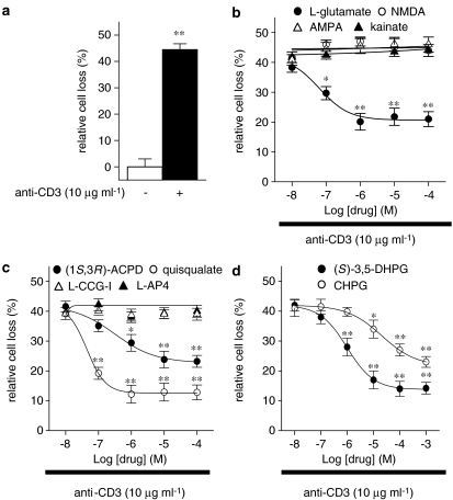
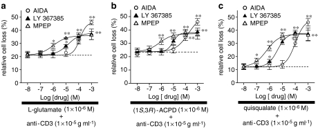
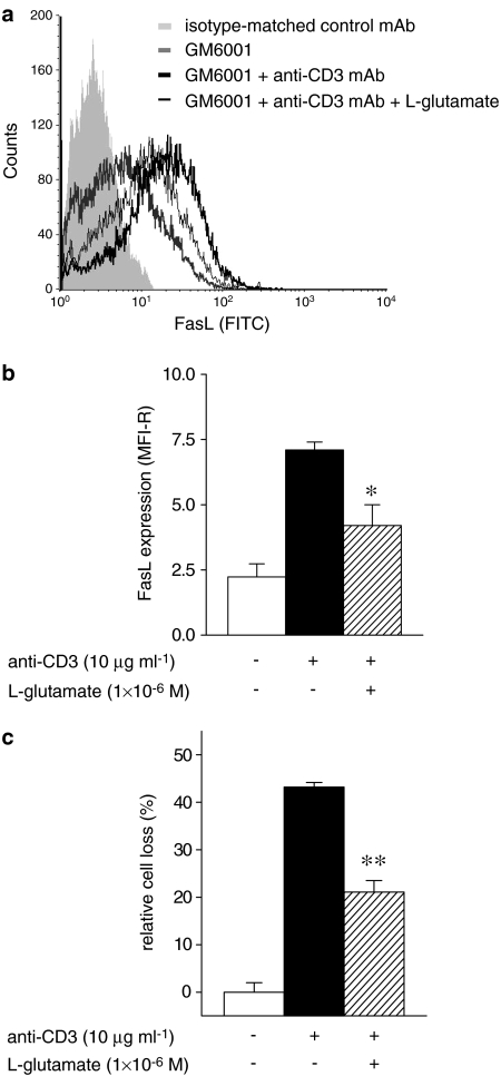
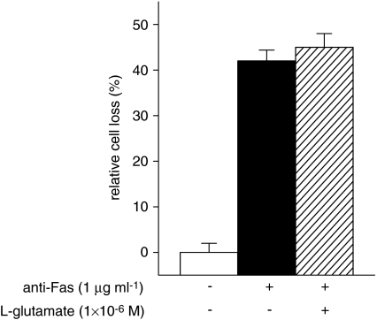
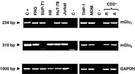
1. The effects of L-glutamate on activation-induced cell death (AICD) of human activated (1 microg ml(-1) phytohemagglutinin plus 2 U ml(-1) interleukin-2; 8 days) T lymphocytes were studied by measuring anti-CD3 monoclonal antibody (10 microg ml(-1); 18 h)-induced cell apoptosis (Annexin V and propidium iodide staining). 2. L-Glutamate (1 x 10(-8)-1 x 10(-4) M) significantly (P < or = 0.01) inhibited AICD in a concentration-dependent manner (EC50=6.3 x 10(-8) M; maximum inhibition 54.8+/-6.3% at 1 x 10(-6) M). 3. The L-glutamate inhibitory effect was pharmacologically characterized as mediated by group I mGlu receptors, since mGlu receptor agonists reproduced this effect. The EC50 values were: 3.2 x 10(-7) M for (1S,3R)-ACPD; 4.5 x 10(-8) M for quisqualate; 1.0 x 10(-6) M for (S)-3,5-DHPG; 2.0 x 10(-5) M for CHPG. 4. Group I mGlu receptor antagonists inhibited the effects of quisqualate 1.0 x 10(-6) M. The IC50 values calculated were: 8.7 x 10(-5), 4.3 x 10(-6) and 6.3 x 10(-7) M for AIDA, LY 367385 and MPEP, respectively. 5. L-Glutamate (1 x 10(-6) M; 18 h) significantly (P < or = 0.05) inhibited FasL expression (40.8+/-11.3%) (cytofluorimetric analysis), whereas it did not affect Fas signalling. 6. Expression of both mGlu1 and mGlu5 receptor mRNA by T lymphocytes and T-cell lines, as demonstrated by reverse transcriptase-PCR analysis, suggests that L-glutamate-mediated inhibition of AICD was exerted on T cells. 7. These data depict a novel role for L-glutamate in the regulation of the immune response through group I mGlu receptor-mediated mechanisms.


Figures

Figure 1
Figure 1
Concentration–response curves for the L-glutamate or iGlu/mGlu receptor agonists inhibitory effects on AICD of human T lymphocytes. Activated T cells were stimulated with anti-CD3 mAb (10 μg ml−1; 18 h) (a); in the presence of increasing concentrations (1 × 10⁻⁸–1 × 10⁻³M) of L-glutamate, NMDA, AMPA or kainate (b), (1S,3R)-ACPD, quisqualate, L-CGG-I or L-AP4 (c), (S)-3,5-DHPG or CHPG (d), and the surviving cells were counted by cytofluorimetric analysis after staining with Annexin V plus propidium iodide. CD3 stimulation induced a 44.5±2.2% of AICD in comparison to anti-CD3-untreated cells. The EC50 values were: 6.3 × 10⁻⁸M for L-glutamate, 3.2 × 10⁻⁷M for (1S,3R)-ACPD, 4.5 × 10⁻⁸M for quisqualate, 1.0 × 10⁻⁶M for (S)-3,5-DHPG and 2.0 × 10⁻⁵M for CHPG. The white and dashed bars show the relative cell loss in anti-CD3-untreated and -treated cells in the absence of other drugs (a). The results are expressed as the mean±s.e.m. of at least ten experiments. *P⩽0.05; **P⩽0.01 versus anti-CD3 mAb-treated cells.
Figure 2
Figure 2
Inhibition of the L-glutamate, (1S,3R)-ACPD or quisqualate protective effects on AICD of human T lymphocytes. Activated T cells were stimulated with anti-CD3 mAb (10 μg ml−1; 18 h) in the presence of (1 × 10⁻⁶M) L-glutamate (a), (1 × 10⁻⁴M) (1S,3R)-ACPD (b), (1 × 10⁻⁶M) quisqualate (c) and increasing concentrations (1 × 10⁻⁸–1 × 10⁻³M) of AIDA, LY 367385 or MPEP. Surviving cells were counted by cytofluorimetric analysis after staining with Annexin V plus propidium iodide. The IC50 values, calculated when quisqualate was used, were: 8.7 × 10⁻⁵M for AIDA; 4.3 × 10⁻⁶M for LY 367385 and 6.3 × 10⁻⁷M for MPEP. Dot lines represent the relative cell loss in anti-CD3 mAb-treated cells in the presence of L-glutamate (1 × 10⁻⁶M) (a), (1S,3R)-ACPD (1 × 10⁻⁴M) (b) quisqualate (1 × 10⁻⁶M) (c). The results are expressed as the mean±s.e.m. of cell death of at least eight experiments. *P⩽0.05, **P⩽0.01 versus anti-CD3 mAb-treated cells in the presence of mGlu receptor agonists.
Figure 3
Figure 3
FasL expression in human T lymphocytes. Activated T cells were stimulated with anti-CD3 mAb (10 μg ml−1; 18 h), in the presence of GM6001 (3 × 10⁻⁵M), and in the absence or presence of L-glutamate (1 × 10⁻⁶M). FasL expression evaluated by direct immunofluorescence and cytofluorimetric analysis using a fluorescein isothiocyanate (FITC)-conjugated anti-FasL mAb (a). (b) FasL expression of L-glutamate (1 × 10⁻⁶M) untreated or treated T cells expressed as MFI-R (see Methods) of total T lymphocytes. Activated T cells were stimulated with anti-CD3 mAb (10 μg ml−1; 18 h) in the presence of GM6001 (3 × 10⁻⁵M) and in the absence or presence of L-glutamate (1 × 10⁻⁶M). Surviving cells were counted by cytofluorimetric analysis after staining with Annexin V plus propidium iodide (c). The results are expressed as the mean±s.e.m. of cell death of at least four experiments. *P⩽0.05, **P⩽0.01 versus anti-CD3 mAb-treated cells in the absence of L-glutamate.
Figure 4
Figure 4
Fas-induced T-cell death. Activated T cells were stimulated with anti-Fas mAb (1 μg ml−1; 18 h), in the absence or presence of L-glutamate (1 × 10⁻⁶M). Surviving cells were counted by cytofluorimetric analysis after staining with Annexin V plus propidium iodide. The results are expressed as the mean±s.e.m. of cell death of at least four experiments.
Figure 5
Figure 5
Expression of mGlu1 and mGlu5 receptors in human lymphoid and myeloid cells. RT–PCRs were performed on total RNA isolated from mouse fibroblasts L(tk) cells stably transfected with human mGlu1 or mGlu5 receptor cDNA (C+), FRO, SUP-T1, H9, HuT-78, Jurkat, THP-1 cell lines, human MDM, human resting T cell (R-CD3+) or human PHA (1 μg ml−1) plus IL-2 (2 U ml−1)-activated T cells (A-CD3+) using specific primers; in the negative control (C-) reverse trascriptase was omitted. GAPDH was used as house keeping gene to normalize cDNA amount. The results are representative of at least five experiments.

References

    1. ALLEN J.W., KNOBLACH S.M., FADEN A.I. Activation of group I metabotropic glutamate receptors reduces neuronal apoptosis but increases necrotic cell death in vitro. Cell Death Differ. 2000;7:470–476. - PubMed
    1. AOUDJIT F., VUORI K. Engagement of the α2β1 integrin inhibits Fas ligand expression and activation-induced cell death in T cells in a focal adhesion kinase-dependent manner. Blood. 2000;95:2044–2051. - PubMed
    1. BALAZS R., MILLER S., CHUN Y., O'TOOLE J., COTMAN C.W. Metabotropic glutamate receptor agonists potentiate cyclic AMP formation induced by forskolin or β-adrenergic receptor activation in cerebral cortical astrocytes in culture. J. Neurochem. 1998;70:2446–2458. - PubMed
    1. BOLDYREV A.A., CARPENTER D.O., JOHNSON P. Emerging evidence for a similar role of glutamate receptors in the nervous and immune systems. J. Neurochem. 2005;95:913–918. - PubMed
    1. BOLDYREV A.A., KAZEY V.I., LEINSOO T.A., MASHKINA A.P., TYULINA O.V., JOHNSON P., TUNEVA J.O., CHITTUR S., CARPENTER D.O. Rodent lymphocytes express functionally active glutamate receptors. Biochem. Biophys. Res. Commun. 2004;324:133–139. - PubMed

Publication types