. 2006 Jun;2(6):e51.
doi: 10.1371/journal.ppat.0020051. Epub 2006 Jun 2.

Envelope exchange for the generation of live-attenuated arenavirus vaccines

Affiliations

Envelope exchange for the generation of live-attenuated arenavirus vaccines

Andreas Bergthaler et al. PLoS Pathog. 2006 Jun.

Abstract

Arenaviruses such as Lassa fever virus cause significant mortality in endemic areas and represent potential bioterrorist weapons. The occurrence of arenaviral hemorrhagic fevers is largely confined to Third World countries with a limited medical infrastructure, and therefore live-attenuated vaccines have long been sought as a method of choice for prevention. Yet their rational design and engineering have been thwarted by technical limitations. In addition, viral genes had not been identified that are needed to cause disease but can be deleted or substituted to generate live-attenuated vaccine strains. Lymphocytic choriomeningitis virus, the prototype arenavirus, induces cell-mediated immunity against Lassa fever virus, but its safety for humans is unclear and untested. Using this virus model, we have developed the necessary methodology to efficiently modify arenavirus genomes and have exploited these techniques to identify an arenaviral Achilles' heel suitable for targeting in vaccine design. Reverse genetic exchange of the viral glycoprotein for foreign glycoproteins created attenuated vaccine strains that remained viable although unable to cause disease in infected mice. This phenotype remained stable even after extensive propagation in immunodeficient hosts. Nevertheless, the engineered viruses induced T cell-mediated immunity protecting against overwhelming systemic infection and severe liver disease upon wild-type virus challenge. Protection was established within 3 to 7 d after immunization and lasted for approximately 300 d. The identification of an arenaviral Achilles' heel demonstrates that the reverse genetic engineering of live-attenuated arenavirus vaccines is feasible. Moreover, our findings offer lymphocytic choriomeningitis virus or other arenaviruses expressing foreign glycoproteins as promising live-attenuated arenavirus vaccine candidates.


Conflict of interest statement

Competing interests. The authors have declared that no competing interests exist.

Figures

Figure 1
Figure 1. Wild-Type and Recombinant Viruses Used in this Study
All LCM viruses used in this study contain an identical L segment encoding for the LCMV RNA-dependent RNA polymerase (L ORF) and for the matrix protein Z. All four viruses also encode for an identical NP translation product. The three partially cDNA derived viruses (rLCMV/INDG, rLCMV/NJG, and rLCMV-ARM*) carry, however, two noncoding genetic tags in the NP ORF as indicated (*; see also Figures 4 and S6). The LCMV-GP ORF of LCMV-ARM and rLCMV-ARM* is substituted for INDG and NJG in rLCMV/INDG and rLCMV/NJG, respectively. Arrows indicate the orientation of individual ORFs. Inverted writing (NP, L) indicates that in the viral genome, the respective ORFs are encoded in antisense polarity.
Figure 2
Figure 2. Attenuation of rLCMV/INDG Prevents Lethal Disease in Infected Mice
(A, B) C57BL/6 mice were infected i.v. with 2 × 10⁴ PFU of either rLCMV/INDG or LCMV-ARM. At the indicated time points after inoculation, groups of two or three mice were killed and viral titers in spleen (A) and liver (B) were determined by immunofocus assay. Symbols represent individual mice. (C) C57BL/6 mice (four per group) were infected with 3 × 10³ PFU rLCMV/INDG or LCMV-ARM i.c. At the time points indicated, they were monitored for clinical signs of choriomeningitis. Animals with terminal disease were killed according to the Swiss law for animal protection. (D) Mice were infected with 3 × 10³ PFU rLCMV/INDG i.c. Ten days later, NP396-specific CD8+ T cells in peripheral blood were enumerated using MHC class I tetramers. Numbers in the upper right quadrants indicate the percentage of NP396-specific CD8+ T cells within the total CD8+ T cell population. Each panel (A–D) shows the results from one representative experiment of two similar ones.
Figure 3
Figure 3. Efficient Control of High-Dose LCMV-WE Infection and Prevention of Liver Disease by rLCMV/INDG-Induced Memory CTL
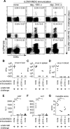
(A) C57BL/6 mice were infected with 2 × 10⁴ PFU rLCMV/INDG i.v. or with 3 × 10³ PFU rLCMV/INDG i.c. on day −109 and on day −314, respectively. On day 0, the immunized mice and a group of naïve control mice (“none”) were challenged with 2 × 10⁵ PFU LCMV-WE i.v. The frequency of NP396-specific memory CD8+ T cells in peripheral blood was assessed prior to challenge (day 0) and on day 4 and day 7 after challenge using MHC class I tetramers. The numbers in the upper right quadrants indicate the frequency of NP396-specific CD8+ T cells within the total CD8+ T cell population in peripheral blood and represent the mean ± SD of three or four mice per group and time point. (B–D) All mice were killed 7 d after challenge, and viral titers in liver, spleen, and blood were determined by immunofocus assay. (E, F) On day 7, serum AST and ALT levels were determined. (G) Liver sections were scored for hepatitis. The symbols in B–G represent individual mice. Statistical analysis was carried out for E–G. Significant differences between groups are indicated for **P < 0.01. All findings described were reproduced in the experiment shown in Figure S5.
Figure 4
Figure 4. Generation and Characterization of rLCMV/NJG
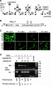
(A) Schematic describing the protocol for generation of rLCMV/NJG. (B) pSNJ(−) was designed analogous to pSIND (also referred to as pSr(−) [36]). It expresses in genomic (−) polarity a recombinant LCMV S segment where the LCMV-GP ORF was substituted for the NJG gene. Transcription is driven by the murine polymerase I promotor (PIP) and is terminated upstream of the murine polymerase I terminator (PIT). UTR, untranslated region; IGR, intergenic region; *noncoding single nucleotide tags. (C) BHK-21 cells on duplicate coverslips were infected for 24 h with supernatants collected from the experiment outlined in A. Staining with INDG- or NJG-specific mAbs as indicated provided a rough estimate of the relative proportions of rLCMV/INDG and rLCMV/NJG contained in the supernatants tested. (D) BHK-21 cells (106 per M6 tissue culture well) were infected with different doses of rLCMV/INDG or of rLCMV/NJG or with both viruses in different combinations as indicated in the chart. At 48 h later, the cells as well as the supernatant were collected. Total cellular RNA was extracted and was processed for detection of INDG and NJG RNA by RT-PCR. The PCR products were separated by agarose gel electrophoresis and were visualized by ethidium bromide staining. Specific amplification products of the expected size are indicated with arrows. Viral infectivity in the supernatant (SN) was measured by immunofocus assay [PFU/ml SN (w/o nAb) listed in the chart under the gel picture]. In addition, an aliquot of supernatant was incubated with VSV-NJ neutralizing mAb H6B9D5 prior to testing the remaining infectivity [see PFU/ml SN (+VSV-NJ nAb)] to discriminate rLCMV/INDG from rLCMV/NJG infectivity.
Figure 5
Figure 5. Attenuated Propagation of rLCMV/NJG but Not of rLCMV-ARM*
(A) BHK-21 cells (106 per M6 well) were infected with the indicated viruses at a multiplicity of infection of 0.01, and infectious virus in the supernatant was measured at the indicated time points. Symbols indicate the mean of three tissue culture wells (SD bars project into the symbol size). (B) BHK-21 cells in M6 tissue culture wells were infected with LCMV-ARM or rLCMV-ARM* at multiplicity of infection of 0.01, and infectious virus in the supernatant was measured at the indicated time points. The symbols represent values from a single cell culture well. One representative experiment of two is shown. (C) C57BL/6 mice infected i.v. with 105 PFU LCMV-ARM or rLCMV-ARM* were killed at the indicated time points, and virus titers in spleen were measured. Symbols represent individual mice. Analogous results were obtained from liver tissue (not shown). One representative experiment of two is shown.

References

    1. Buchmeier MJ, Bowen MD, Peters CJ. Arenaviridae: The viruses and their replication. In Knipe DM, editor. Fields Virology. Philadelphia: Lippincott Williams & Wilkins; 2001. pp. 1635–1668.
    1. Perez M, Craven RC, De La Torre JC. The small RING finger protein Z drives arenavirus budding: Implications for antiviral strategies. Proc Natl Acad Sci U S A. 2003;100:12978–12983. - PMC - PubMed
    1. McCormick JB, Webb PA, Krebs JW, Johnson KM, Smith ES. A prospective study of the epidemiology and ecology of Lassa fever. J Infect Dis. 1987;155:437–444. - PubMed
    1. Borio L, Inglesby T, Peters CJ, Schmaljohn AL, Hughes JM, et al. Hemorrhagic fever viruses as biological weapons: Medical and public health management. JAMA. 2002;287:2391–2405. - PubMed
    1. Charrel RN, de Lamballerie X. Arenaviruses other than Lassa virus. Antiviral Res. 2003;57:89–100. - PubMed

Publication types

MeSH terms