. 2006 Jun 12;203(6):1603-14.
doi: 10.1084/jem.20052543. Epub 2006 Jun 5.

Nepmucin, a novel HEV sialomucin, mediates L-selectin-dependent lymphocyte rolling and promotes lymphocyte adhesion under flow

Affiliations

Nepmucin, a novel HEV sialomucin, mediates L-selectin-dependent lymphocyte rolling and promotes lymphocyte adhesion under flow

Eiji Umemoto et al. J Exp Med. .

Abstract

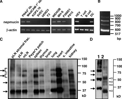
Lymphocyte trafficking to lymph nodes (LNs) is initiated by the interaction between lymphocyte L-selectin and certain sialomucins, collectively termed peripheral node addressin (PNAd), carrying specific carbohydrates expressed by LN high endothelial venules (HEVs). Here, we identified a novel HEV-associated sialomucin, nepmucin (mucin not expressed in Peyer's patches [PPs]), that is expressed in LN HEVs but not detectable in PP HEVs at the protein level. Unlike conventional sialomucins, nepmucin contains a single V-type immunoglobulin (Ig) domain and a mucin-like domain. Using materials affinity-purified from LN lysates with soluble L-selectin, we found that two higher molecular weight species of nepmucin (75 and 95 kD) were decorated with oligosaccharides that bind L-selectin as well as an HEV-specific MECA-79 monoclonal antibody. Electron microscopic analysis showed that nepmucin accumulates in the extended luminal microvillus processes of LN HEVs. Upon appropriate glycosylation, nepmucin supported lymphocyte rolling via its mucin-like domain under physiological flow conditions. Furthermore, unlike most other sialomucins, nepmucin bound lymphocytes via its Ig domain, apparently independently of lymphocyte function-associated antigen 1 and very late antigen 4, and promoted shear-resistant lymphocyte binding in combination with intercellular adhesion molecule 1. Collectively, these results suggest that nepmucin may serve as a dual-functioning PNAd in LN HEVs, mediating both lymphocyte rolling and binding via different functional domains.


Figures

Figure 1.
Figure 1.
Amino acid sequence of mouse nepmucin and putative exon/intron structure. (A) The deduced amino acid sequence of nepmucin. The putative signal sequence and the transmembrane region are represented by a broken underline and double underline, respectively. The putative Ig domain is underlined, and the cysteine residues participating in the Ig domain structure are circled. The tandem repeats in the mucin-like domain are boxed. (B) The exon/intron structure of mouse nepmucin gene and splicing isoforms. The genomic structure of nepmucin was deduced by comparing the genomic sequence (AL591145) with the cDNA sequence of each nepmucin isoform (isoforms A, B, C, and D). All intron-exon boundaries follow the AG/GT rules. The nucleotide sequences of the nepmucin isoforms have been submitted under accession numbers AB243063, AB243064, AB243065, and AB243066. TM, transmembrane domain.
Figure 2.
Figure 2.
Cell and tissue expression of nepmucin. (A) Nepmucin mRNA expression analyzed by RT-PCR. Left: HEV cDNA libraries (PNAd+ HEVs and MAdCAM-1+ HEVs), endothelial cell lines (KOP2.16, SVEC4-10, bEND3, and F2), monocyte/macrophage cell lines (WEHI3B, MH-S, and P388D1), and a mast cell line (P815) were analyzed. Right: Freshly isolated MAdCAM-1+ HEV cells, T cells, B cells, and DCs were analyzed. The PCR primers were designed to detect all the nepmucin isoforms as a single band. (B) Expression of nepmucin splicing isoforms in freshly isolated MAdCAM-1+ HEVs. A primer pair designed to detect nepmucin isoforms with different product sizes was used. (C) Tissue distribution of nepmucin. The expression of nepmucin was analyzed by Western blotting using the anti-nepmucin mAb ZAQ2, which recognized all four nepmucin variants (arrows). Control rat IgG2a did not give any specific signals (unpublished data). (D) Enzymatic O-deglycosylation of nepmucin. Lysates from mouse heart were subjected to immunoprecipitation with ZAQ2 and mock-treated (lane 1) or treated with O-glycanase (lane 2). The proteins were analyzed with the anti-nepmucin mAb, ZAQ3. The arrows and arrowheads indicate the four kinds of antigenic components.
Figure 3.
Figure 3.
Distribution pattern of nepmucin in mouse tissues. Two-color immunostaining of peripheral LNs with anti-nepmucin (A), anti-PNAd (B), and the merged image (C); two-color immunostaining of mesenteric LNs with anti-nepmucin (D), anti-MAdCAM-1 (E), and the merged image (F). A–F shows that nepmucin was expressed in HEVs in peripheral LNs and mesenteric LNs. Two-color staining of PPs with anti-nepmucin (G) and anti–MAdCAM-1 (H) revealed that nepmucin was undetectable in PP HEVs. Double staining with anti-nepmucin and anti-CD31 showed that nepmucin was also undetectable in non–HEV-type vessels in PPs (I). Two color staining of spleen with anti-nepmucin (J–L) and anti-CD31 (J and K) or anti–MAdCAM-1 (L). The expression of nepmucin was found in the marginal sinus and trabecula (J), but not in central arteries (K). Nepmucin was also detected in the MAdCAM-1+ sinus-lining cells (L). Two-color staining of heart with anti-nepmucin (M), anti-CD31 (N), and the merged image (O) showed that nepmucin was selectively expressed in the intra-muscular capillaries but not the arteries. Scale bar, 50 μm (C, F, K, L, and O) and 100 μm (H, I, and J). Negative control rat IgG for anti-nepmucin did not bind tissue sections (unpublished data).
Figure 4.
Figure 4.
Interaction of nepmucin expressed in peripheral LN HEVs with L-selectin and MECA-79 mAb. (A) Reactivity of L-selectin–binding materials with anti-nepmucin mAb. Total cell lysates of peripheral LNs were immunoblotted with the anti-nepmucin mAb ZAQ2 (left). Lysates of peripheral LNs were precipitated with LEC/IgG chimeric protein or control human IgG, with or without EDTA, and analyzed by Western blotting with ZAQ2 (right). The two major antigenic components are indicated by arrowheads. (B) Reactivity of the nepmucin in peripheral LN HEVs with the MECA-79 mAb. Total cell lysates of peripheral LNs were immunoblotted with the MECA-79 mAb (left). Lysates of mouse peripheral LNs were immunoprecipitated with anti-nepmucin or control rat IgG, and immunoblotted with MECA-79 (middle) or the anti-nepmucin mAb ZAQ2 (right). MECA-79 recognized three nepmucin species (arrows) among the four isoforms (white arrowheads). In A and B, isotype-matched controls to anti-nepmucin and MECA-79 gave no specific signals (unpublished data). (C) Reprecipitation of nepmucin from L-selectin–binding materials. L-selectin–binding materials were reprecipitated with ZAQ2 or rat IgG and analyzed by Western blotting using MECA-79 (left) or ZAQ2 (right). (D) Sensitivity of nepmucin to OSGE or sialydase. L-selectin–binding materials treated with OSGE were subjected to immunoprecipitation with the anti-nepmucin mAb, ZAQ2 (left). Alternatively, L-selectin–binding materials were reprecipitated with ZAQ2 and then treated with sialydase (right). The precipitates were analyzed with MECA-79. Arrowheads indicate the higher molecular weight species of nepmucin.
Figure 5.
Figure 5.
Ultrastructural localization of nepmucin in peripheral LN HEVs. (A) Anti-nepmucin mAb ZAQ5 bound to the vascular luminal surface of LN HEVs. (B) A higher magnification view of the area shown within the square in A shows that nepmucin was frequently concentrated on the extended luminal microvillous process near the intracellular junction. In addition, nepmucin appeared to be localized in the site of internalized plasma membrane (arrow) and in the tubular- or spherical-shaped endosomal vesicles (arrowheads) below the endothelial cell surface. (C) Nepmucin was detectable on the lateral surface of LN HEVs. A multivesicular body (asterisk) was positive for nepmucin. (D) A higher magnification view of the lateral junction area shown within the square in C showed the anti-nepmucin mAb located on the lateral membranes of the high endothelial cells, where they formed loose connections (arrowheads). Ly, lymphocyte. Bars, 1 μm (A and C) and 0.5 μm (B and D).
Figure 6.
Figure 6.
L-selectin–dependent cell rolling mediated by nepmucin-Fc produced in CHO cells expressing C2GnT, C1GnT, FucTVII, and LSST. (A) Preparation of chimeric proteins decorated with MECA-79+ sugar chains. Nepmucin FL-Fc, ΔIg-Fc, Δmucin-Fc, and GlyCAM-1-Fc were produced in the A5-Core1 cells and analyzed with MECA-79 mAb. (B) Lymphocyte rolling on immobilized chimeras under flow. The inside wall of capillary tubes was coated with one of the chimeric proteins or human IgG (20 μg/ml). Jurkat cells (2 × 10⁶ cells/ml) were infused into the capillary tubes (0.8 dyne/cm2), and the rolling cell number was determined. (C) Lymphocyte rolling velocity at a shear stress of 0.8 dyne/cm2. The histograms display the rolling cell number observed at the indicated rolling velocity. (D) Inhibition of lymphocyte rolling by EDTA or anti– L-selectin mAb. Lymphocytes were pretreated with EDTA, anti–L-selectin (DREG-56), or control human IgG1 before the infusion. (E) Inhibition of lymphocyte rolling by treating immobilized nepmucin with MECA-79, OSGE, or sialydase. Glass capillaries were first coated with Fc chimeras and treated as indicated before the rolling assay.
Figure 7.
Figure 7.
Lymphocyte binding to nepmucin via its Ig domain. (A) Binding of lymphocytes to nepmucin-Fc chimeras. Nepmucin FL-Fc, ΔIg-Fc, Δmucin-Fc, GlyCAM-1-Fc, ICAM-1-Fc, and human IgG1 Fc were immobilized on glass slides and incubated with splenocytes in the presence or absence of PMA. The number of cells bound per unit area was determined microscopically, and the mean ± SD from three independent areas is given. The data are representative of at least three independent experiments. (B) Effects of anti-nepmucin mAb pretreatment on lymphocyte adhesion. Coated proteins were incubated with a panel of anti-nepmucin mAbs that specifically recognize the Ig domain (ZAQ1, ZAQ2, ZAQ3, and ZAQ4) or with the control rat IgG. The number of cells bound under PMA stimulation is shown. (C) The effects of anti–LFA-1 and/or anti–VLA-4 on nepmucin-mediated lymphocyte binding. Lymphocytes pretreated with anti–LFA-1 and/or anti–VLA-4 or rat IgG were incubated with or without PMA. (D) Effects of EDTA and Mn2+ on nepmucin-mediated lymphocyte binding. Splenocytes were preincubated in the presence or absence of EDTA on immobilized proteins and stimulated with or without PMA or MnCl2. (E) Detachment of lymphocytes bound to nepmucin under flow. Fc chimeras were immobilized on the inner surface of glass capillaries with or without CCL21. Lymphocytes were injected into the capillaries and allowed to accumulate on the substrate at a shear stress of 0.125 dyne/cm2. The shear stress was then increased in twofold increments every 20 s. The lymphocytes that remained adherent at each shear-stress level were counted and expressed as a percentage of the initially bound cells.
Figure 8.
Figure 8.
Enhancement of lymphocyte adhesion to an ICAM-1–coated substrate by nepmucin under flow conditions. (A) Lymphocyte rolling on Fc chimeras under flow conditions. Nepmucin FL-Fc, GlyCAM-1-Fc, ICAM-1-Fc, or human IgG was immobilized on the inside wall of capillary tubes that had been coated with goat anti–human IgG. Lymphocytes were infused into the capillary tubes (1.1 dyne/cm2), and the number of cells that rolled in 0.64 mm2 per min was counted. (B) Lymphocyte adhesion on nepmucin FL-Fc coimmobilized with ICAM-1-Fc under flow conditions. Fc chimeric proteins were coimmobilized on the inside of capillary tubes with or without CCL21 as indicated. Lymphocytes were infused into the capillary, and the number of bound cells in 0.29 mm2 was determined. Data represent the mean ± SD from three independent areas. *, P < 0.005. (C) Lymphocyte adhesion to nepmucin's Ig domain under flow. Fc chimeras and CCL21 were immobilized on the inside of capillaries as in B. Lymphocytes were injected into the capillaries, and bound cells were counted.

References

    1. von Andrian, U.H., and T.R. Mempel. 2003. Homing and cellular traffic in lymph nodes. Nat. Rev. Immunol. 3:867–878. - PubMed
    1. Miyasaka, M., and T. Tanaka. 2004. Lymphocyte trafficking across high endothelial venules: dogmas and enigmas. Nat. Rev. Immunol. 4:360–370. - PubMed
    1. Rosen, S.D. 2004. Ligands for L-selectin: homing, inflammation, and beyond. Annu. Rev. Immunol. 22:129–156. - PubMed
    1. Yeh, J.C., N. Hiraoka, B. Petryniak, J. Nakayama, L.G. Ellies, D. Rabuka, O. Hindsgaul, J.D. Marth, J.B. Lowe, and M. Fukuda. 2001. Novel sulfated lymphocyte homing receptors and their control by a Core1 extension β1,3-N-acetylglucosaminyltransferase. Cell. 105:957–969. - PubMed
    1. Maly, P., A. Thall, B. Petryniak, C.E. Rogers, P.L. Smith, R.M. Marks, R.J. Kelly, K.M. Gersten, G. Cheng, T.L. Saunders, et al. 1996. The α(1,3)fucosyltransferase Fuc-TVII controls leukocyte trafficking through an essential role in L-, E-, and P-selectin ligand biosynthesis. Cell. 86:643–653. - PubMed

Publication types