. 2006 Jun 12;203(6):1591-601.
doi: 10.1084/jem.20060905. Epub 2006 Jun 5.

Yersinia pseudotuberculosis disseminates directly from a replicating bacterial pool in the intestine

Affiliations

Yersinia pseudotuberculosis disseminates directly from a replicating bacterial pool in the intestine

Penelope D Barnes et al. J Exp Med. .

Erratum in

  • J Exp Med. 2006 Jul 10;203(7):1829

Abstract

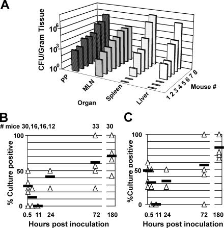
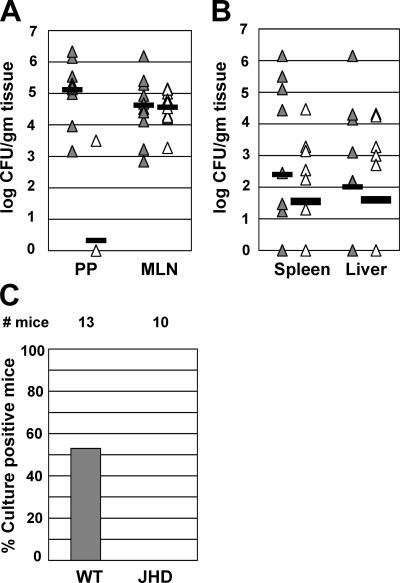
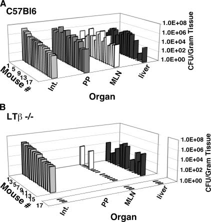
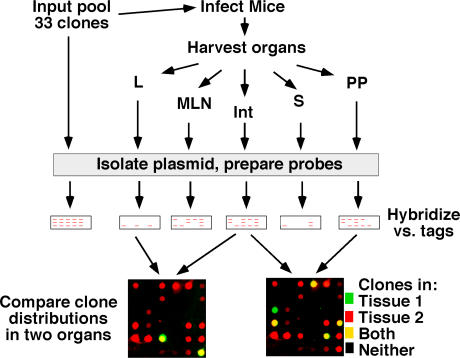
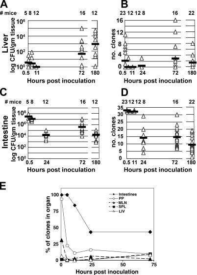
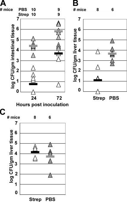
Dissemination of Yersinia pseudotuberculosis within mice after oral inoculation was analyzed. Y. pseudotuberculosis translocated to organs such as the liver and spleen shortly after oral inoculation, but was quickly cleared. In contrast, a second temporally distinct bacterial translocation event resulted in successful hepatosplenic replication of the bacteria. Replicating pools of bacteria could be established in these organs in mouse mutants that lacked Peyer's patches. These animals frequently had sterile mesenteric lymph nodes, a finding consistent with translocation taking place independently of regional lymph node colonization. In further contradiction to accepted models for dissemination of enteropathogens, clonal analysis revealed that bacteria causing disease in the spleen and liver of C57BL/6J mice were derived from populations located outside the intestinal lymph nodes. Replication of bacteria in the intestine before translocation appeared critical for dissemination, as transient selective suppression by streptomycin of bacterial growth in the intestine delayed dissemination of Y. pseudotuberculosis. These results collectively indicate that hepatosplenic colonization appears intimately connected with the ability of Y. pseudotuberculosis to successfully establish replication in the intestinal lumen and does not result from ordered spread leading from the intestine to regional lymph nodes before dissemination.


Figures

Figure 1.
Figure 1.
Typical infection of C57BL/6J mice 7 d after orogastric inoculation of Y. pseudotuberculosis and temporal kinetics of hepatosplenic colonization. C57BL/6J mice were orogastrically inoculated with 5 × 10⁸ CFU of Y. pseudotuberculosis. 7 d after infection (A), the mice were killed and the organs were weighed and cultured for bacterial colony counts on MacConkey lactose plates (see Materials and methods). Similar results were obtained with BALB/c mice (see Fig. 2 A). PP, Peyer's patches; MLN, mesenteric lymph nodes. After oral inoculation, at 0.5, 5, 11, 24, 72, and 180 h after inoculation, groups of mice were killed and the spleens (B) and livers (C) were cultured for the presence of any bacteria using the broth recovery technique (see Materials and methods). The results, compiled from a series of experiments and displayed above the graph in B, are the total number of mice examined at each time point. Each triangle represents the percentage of mice with bacteria cultured from spleen or liver in one experiment at each time point. Bars are the mean percentages of organs containing bacteria. At some time points, the same proportion was obtained in different experiments (see 11 h); consequently, the triangles are superimposed. The total number of experiments performed at each time point were as follows: 0.5 h, seven experiments; 5 h, four experiments; 11 h, four experiments; 24 h, three experiments; 72 h, seven experiments; and 180 h, seven experiments.
Figure 2.
Figure 2.
Oral infection by Y. pseudotuberculosis in B cell–deficient mice indicates two mechanistically different waves of spread from the intestine. 6 d after oral inoculation with 5 × 10⁸ CFU of Y. pseudotuberculosis (see Materials and methods), BALB/c mice and BALB/c-JHD mice (B cell deficient) were killed and (A) PP and MLN or (B) spleen and liver were cultured for colony counts (see Materials and methods). Each triangle represents the CFU recovered from the organ of a mouse: (closed triangles) BALB/c mice and (open triangles) BALB/c-JHD mice. At some time points, similar colony counts were obtained in different mice and the triangles are superimposed. Bars are the mean number of bacteria found in the tissues from all experiments. The results were accumulated from three separate experiments. In total, 10 wild-type and 11 BALB/c-JHD mice were analyzed. There was no statistical difference in CFU recovered between the two mouse strains from the liver, spleen, or MLN. Only three BALB/c-JHD mice contained identifiable PP and bacteria were cultured from the PP of only one of these mice. (C) B cell function is required for early dissemination of Y. pseudotuberculosis from the intestine to spleen and liver. BALB/c and BALB/c-JHD mice were orogastrically inoculated with 5 × 10⁸ CFU of Y. pseudotuberculosis. The mice were killed between 30 min and 2 h after infection and the liver and spleens were cultured for the presence of bacteria, using the broth recovery technique. The results are pooled from three separate experiments. # mice, mean percentage of mice from three separate experiments that had culturable bacteria from the liver or spleen in the noted mouse strain.
Figure 3.
Figure 3.
Y. pseudotuberculosis colonizes the livers of the LT β−/− mouse. Mice were orally inoculated with 5 × 10⁸ Y. pseudotuberculosis. 6 d after inoculation, they were killed and colony counts were obtained from noted tissues. Shown are individual animals in which intestinal colonization was established. Shown are three experiments from (A) C57BL/6J mice and (B) LT β−/− mice. The LT β−/− mice are displayed as separate graphs, with the three mice showing no intestinal bacteria separated from the other data. No PPs were found in the LT β−/− mouse, whereas only two mice had detectable colonization of the MLN in the KO strain.
Figure 4.
Figure 4.
Strategy for identification of Y. pseudotuberculosis clones present in different tissue sites. 33 Y. pseudotuberculosis strains, each differing by only a unique oligonucleotide tag, were grown in broth and pooled immediately before oral inoculation into C57BL/6J mice (see Materials and methods). At various times after inoculation, the animals were killed, noted glands were removed, and bacteria were isolated by plating tissues on bacteriologic media (see Materials and methods). The colonies arising were pooled, DNA was isolated, and probes were constructed by PCR amplification of the unique oligonucleotide tags. The clones found in each tissue site were determined, and compared with the results obtained from other tissue sites.
Figure 5.
Figure 5.
Bacterial load and clonal dissemination of Y. pseudotuberculosis. C57BL/6J mice were orally inoculated with 5 × 10⁸ CFU of a pool of 33 uniquely tagged wild-type Y. pseudotuberculosis clones. At noted times after inoculation, the mice were killed and the liver (A and B) or intestines (C and D) (see Materials and methods for “intestines” definition) were assayed for the bacterial CFU (A and C) or the number of individual clones (B and D) (see Materials and methods). The results are pooled from separate experiments. Displayed above the graphs are the total number of mice examined at each time point and the number of experiments performed for each time point. Each triangle represents the CFU or number of clones isolated from the liver or intestines of one mouse in one experiment at each time point. Dark bars are the mean CFU or clones isolated from the livers of all mice at that time point. At some time points, the same CFU or number of clones was obtained in different mice; consequently, some triangles are superimposed. (E) Clonal distribution in infected organs over time. To determine the percentage of total clones found in each tissue, the total number of clones found in each tissue site was determined for all the animals killed at the noted time point. This number was then divided by the total number of clones that had been inoculated in all the animals analyzed at the noted time point to obtain the percentage.
Figure 6.
Figure 6.
Oral streptomycin reduces intestinal bacterial load and subsequent systemic infection after oral but not intraperitoneal inoculation. C57BL/6J mice were orally inoculated (A and B) with 5 × 10⁸ CFU of Y. pseudotuberculosis or inoculated i.p. (C) with 104 CFU of Y. pseudotuberculosis. At 5, 12, and 24 h after inoculation, each mouse was orogastrically inoculated either with streptomycin (white triangles and black bars) or with PBS (gray triangles and bars). 24 and 72 h after inoculation, the intestines (see Materials and methods) were examined for CFU (A). The livers of the orally inoculated mice were also examined for colony counts 72 h after infection (B). The mice inoculated i.p. with Y. pseudotuberculosis were killed 72 h after inoculation and examined for CFU (C). The results are pooled from three separate experiments. Displayed above the graph is the total number of mice examined at each time point. Each triangle represents the CFU of a mouse. Bars are the mean number of bacteria found in the tissues from all experiments. At some time points, similar colony counts were obtained in different mice and the triangles are superimposed.

References

    1. Achtman, M., K. Zurth, G. Morelli, G. Torrea, A. Guiyoule, and E. Carniel. 1999. Yersinia pestis, the cause of plague, is a recently emerged clone of Yersinia pseudotuberculosis. Proc. Natl. Acad. Sci. USA. 96:14043–14048. - PMC - PubMed
    1. Naktin, J., and K.G. Beavis. 1999. Yersinia enterocolitica and Yersinia pseudotuberculosis. Clin. Lab. Med. 19:523–536. - PubMed
    1. Cornelis, G.R. 2000. Molecular and cell biology aspects of plague. Proc. Natl. Acad. Sci. USA. 97:8778–8783. - PMC - PubMed
    1. Bottone, E.J. 1999. Yersinia enterocolitica: overview and epidemiologic correlates. Microbes Infect. 1:323–333. - PubMed
    1. Hubbert, W.T., C.W. Petenyi, L.A. Glasgow, C.T. Uyeda, and S.A. Creighton. 1971. Yersinia pseudotuberculosis infection in the United States. Septicema, appendicitis, and mesenteric lymphadenitis. Am. J. Trop. Med. Hyg. 20:679–684. - PubMed

Publication types

MeSH terms