. 2006 Jun 13;103(24):9190-5.
doi: 10.1073/pnas.0603503103. Epub 2006 Jun 5.

Therapy-induced antibodies to MHC class I chain-related protein A antagonize immune suppression and stimulate antitumor cytotoxicity

Affiliations

Therapy-induced antibodies to MHC class I chain-related protein A antagonize immune suppression and stimulate antitumor cytotoxicity

Masahisa Jinushi et al. Proc Natl Acad Sci U S A. .

Abstract

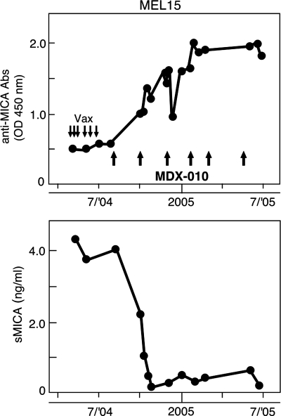
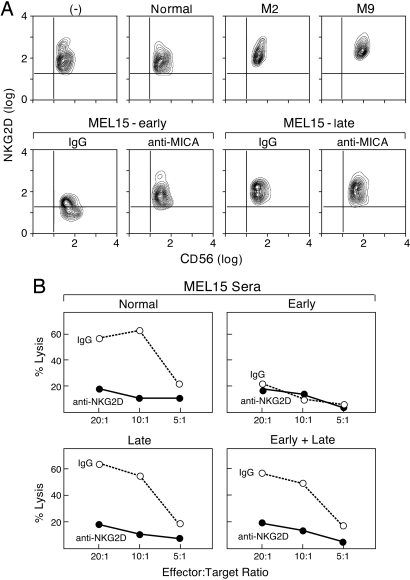
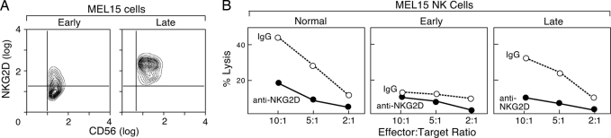
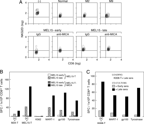
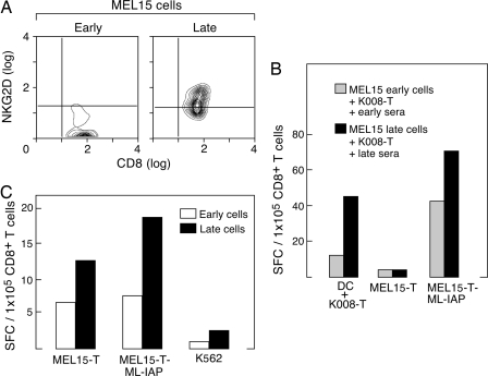
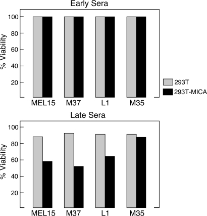
The activation of NKG2D on innate and adaptive cytotoxic lymphocytes contributes to immune-mediated tumor destruction. Nonetheless, tumor cell shedding of NKG2D ligands, such as MHC class I chain-related protein A (MICA), results in immune suppression through down-regulation of NKG2D surface expression. Here we show that some patients who respond to antibody-blockade of cytotoxic T lymphocyte-associated antigen 4 or vaccination with lethally irradiated, autologous tumor cells engineered to secrete granulocyte-macrophage colony-stimulating factor generate high titer antibodies against MICA. These humoral reactions are associated with a reduction of circulating soluble MICA (sMICA) and an augmentation of natural killer (NK) cell and CD8(+) T lymphocyte cytotoxicity. The immunotherapy-induced anti-MICA antibodies efficiently opsonize cancer cells for dendritic cell cross-presentation, which is correlated with a diversification of tumor antigen recognition. The anti-MICA antibodies also accomplish tumor cell lysis through complement fixation. Together, these findings establish a key role for the NKG2D pathway in the clinical activity of cytotoxic T lymphocyte-associated antigen 4 antibody blockade and granulocyte-macrophage colony-stimulating factor secreting tumor cell vaccines. Moreover, these results highlight the therapeutic potential of anti-MICA antibodies to overcome immune suppression and effectuate tumor destruction in patients.


Conflict of interest statement

Conflict of interest statement: No conflicts declared.

Figures

Fig. 1.
Fig. 1.
CTLA-4 blockade after autologous tumor vaccination elicited a potent humoral reaction to MICA that was temporally associated with a reduction in sMICA. (Upper) Longitudinal sera samples from MEL15 were diluted 1:500 and analyzed by ELISA with recombinant MICA protein. Reactivity was determined with a pan-IgG secondary. Downward arrows denote tumor cell vaccinations and upward arrows depict infusions of MDX-010. (Lower) Serial sMICA levels were measured with a sandwich ELISA.
Fig. 2.
Fig. 2.
Therapy-induced anti-MICA antibodies antagonized sMICA suppression of innate immune responses. (A) Normal donor PBMCs were incubated for 48 h in sera, and NKG2D expression on gated NK cells (CD56+ and CD3) was determined with flow cytometry. Sera samples were obtained from a healthy donor, vaccinated melanoma patients without sMICA (M2 and M9), and MEL15 during immunization or after CTLA-4 antibody blockade. Anti-MICA monoclonal antibodies or isotype controls were added where indicated. (B) Healthy donor PBMCs were incubated in normal or MEL15 sera for 48 h and washed; NK cells were then purified with magnetic bead selection and tested for lytic activity against 51Cr-labeled K562 targets. The NKG2D-dependent lysis was determined with the addition of the mAb 1D11 (anti-NKG2D) or isotype control (IgG).
Fig. 3.
Fig. 3.
Immunotherapy restored protective antitumor innate responses in MEL15. (A) PBMCs were obtained from MEL15 during vaccination or after CTLA-4 blockade, and NKG2D expression on gated NK cells was determined with flow cytometry. (B) Magnetic bead-purified healthy donor or MEL15 NK cells obtained at different times were tested for lytic activity against 51Cr-labeled K562 targets.
Fig. 4.
Fig. 4.
Therapy-induced anti-MICA antibodies antagonized sMICA suppression of adaptive immune responses and enhanced MICA-dependent cross-presentation. (A) Normal donor PBMCs were incubated for 48 h in sera, and NKG2D expression on gated CD8+ T cells (CD8+ and CD3+) was determined with flow cytometry. Sera samples were obtained from a healthy donor, vaccinated melanoma patients without sMICA (M2 and M9), and MEL15 during immunization or after CTLA-4 antibody blockade. Anti-MICA monoclonal antibodies or isotype controls were added where indicated. (B) Dendritic cells were generated from adherent PBMCs of HLA-A2.1+ donors, loaded with MEL15 early or late sera-coated MEL15-T or MEL15-T-MICA tumor cells, matured with LPS, and used to stimulate autologous purified CD8+ T cells for 7 days. IFN-γ production was measured by ELISPOT against the indicated targets. No reactivity against unpulsed dendritic cells or dendritic cells loaded with unopsonized tumors was observed (data not shown). (C) HLA-A2.1+ dendritic cells were loaded with MEL15 late sera-coated K008-T melanoma cells (MICA+), LPS matured, and used to stimulate autologous CD8+ T cells in the presence of early or late MEL15 sera. IFN-γ production was measured by ELISPOT against the indicated targets.
Fig. 5.
Fig. 5.
Immunotherapy restored protective antitumor innate responses and enhanced cross-presentation in MEL15. (A) PBMCs were obtained from MEL15 during vaccination or after CTLA-4 blockade, and NKG2D expression on gated CD8+ T cells was determined with flow cytometry. (B) PBMCs were obtained from MEL15 during vaccination or after CTLA-4 blockade and used to generate dendritic cells. These cells were loaded with K008-T melanoma cells coated in early or late MEL15 sera and then matured with LPS. Purified CD8+ T cells from the same time points were then stimulated in vitro with the respective tumor-loaded dendritic cells for 7 days. IFN-γ production was measured by ELISPOT against the indicated targets. (C) MEL15 CD8+ T cells were purified from PBMCs collected during vaccination or after CTLA-4 blockade and tested for IFN-γ production against the indicated targets without prior in vitro stimulation. Melanoma inhibitor of apoptosis protein (ML-IAP) is a previously characterized tumor rejection antigen (13).
Fig. 6.
Fig. 6.
Immunotherapy-induced anti-MICA antibodies mediate complement-dependent lysis. 293T and 293T-MICA embryonic kidney cells were incubated in patient sera and complement, and cell viability was determined with a 3-(4,5-dimethylthiazol-2-yl)-2,5-diphenyl tetrazolium bromide (MTT) assay after 6 h. (Upper) Prevaccination sera. (Lower) Postvaccination sera. M35 was a vaccinated, long-term surviving metastatic melanoma patient (7 years) without anti-MICA antibodies. The differences were abrogated by heat inactivation of the complement.

References

    1. Dunn G. P., Old L. J., Schreiber R. D. Immunity. 2004;21:137–148. - PubMed
    1. Sato E., Olson S. H., Ahn J., Bundy B., Nishikawa H., Qian F., Jungbluth A. A., Frosina D., Gnjatic S., Ambrosone C., et al. Proc. Natl. Acad. Sci. USA. 2005;102:18538–18543. - PMC - PubMed
    1. Dranoff G. Nat. Rev. Cancer. 2004;4:11–22. - PubMed
    1. Hodi F. S., Dranoff G. Adv. Immunol. 2006;90:337–360. - PubMed
    1. Korman A., Peggs K., Allison J. P. Adv. Immunol. 2006;90:293–335. - PMC - PubMed

Publication types

MeSH terms