

Synergic effect of polymorphisms in ERCC6 5' flanking region and complement factor H on age-related macular degeneration predisposition
- PMID: 16754848
- PMCID: PMC1474016
- DOI: 10.1073/pnas.0603485103
Synergic effect of polymorphisms in ERCC6 5' flanking region and complement factor H on age-related macular degeneration predisposition
Abstract
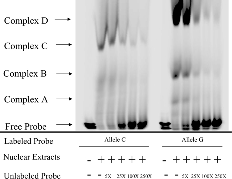
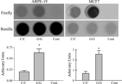
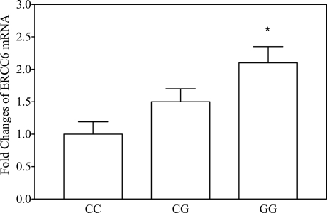
This study investigates age-related macular degeneration (AMD) genetic risk factors through identification of a functional single-nucleotide polymorphism (SNP) and its disease association. We chose ERCC6 because of its roles in the aging process, DNA repair, and ocular degeneration from the gene disruption. Bioinformatics indicated a putative binding-element alteration on the sequence containing C-6530>G SNP in the 5' flanking region of ERCC6 from Sp1 on the C allele to SP1, GATA-1, and OCT-1 on the G allele. Electrophoretic mobility shift assays displayed distinctive C and G allele-binding patterns to nuclear proteins. Luciferase expression was higher in the vector construct containing the G allele than that containing the C allele. A cohort of 460 advanced AMD cases and 269 age-matched controls was examined along with pathologically diagnosed 57 AMD and 18 age-matched non-AMD archived cases. ERCC6 C-6530>G was associated with AMD susceptibility, both independently and through interaction with an SNP (rs380390) in the complement factor H (CFH) intron reported to be highly associated with AMD. A disease odds ratio of 23 was conferred by homozygozity for risk alleles at both ERCC6 and CFH compared with homozygozity for nonrisk alleles. Enhanced ERCC6 expression was observed in lymphocytes from healthy donors bearing ERCC6 C-6530>G alleles. Intense immunostaining of ERCC6 was also found in AMD eyes from ERCC6 C-6530>G carriers. The strong AMD predisposition conferred by the ERCC6 and CFH SNPs may result from biological epistasis, because ERCC6 functions in universal transcription as a component of RNA pol I transcription complex.
Conflict of interest statement
Conflict of interest statement: No conflicts declared.
Figures

References
-
- Klein R., Peto T., Bird A., Vannewkirk M. R. Am. J. Ophthalmol. 2004;137:486–495. - PubMed
-
- Friedman D. S., O’Colmain B. J., Munoz B., Tomany S. C., McCarty C., de Jong P. T., Nemesure B., Mitchell P., Kempen J. Eye Diseases Prevalence Research Group. Arch. Ophthalmol. 2004;122:564–572. - PubMed
-
- Hyman L., Neborsky R. Curr. Opin. Ophthalmol. 2002;13:171–175. - PubMed
Publication types
MeSH terms
Substances
Grants and funding
LinkOut - more resources
Full Text Sources
Medical
Molecular Biology Databases
Miscellaneous
