. 2006 Jun 13;103(24):9045-50.
doi: 10.1073/pnas.0603318103. Epub 2006 Jun 5.

Compartmentalized signaling of Ras in fission yeast

Affiliations

Compartmentalized signaling of Ras in fission yeast

Brian Onken et al. Proc Natl Acad Sci U S A. .

Abstract

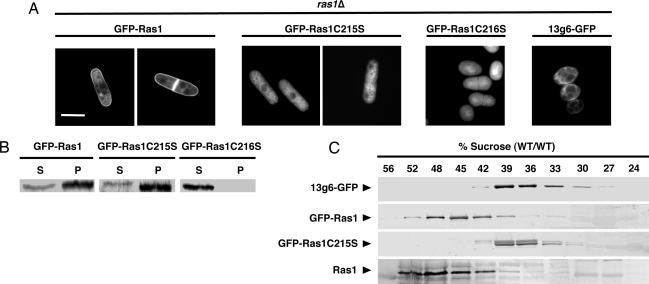
Compartment-specific Ras signaling is an emerging paradigm that may explain the multiplex outputs from a single GTPase. The fission yeast, Schizosaccharomyces pombe, affords a simple system in which to study Ras signaling because it has a single Ras protein, Ras1, that regulates two distinct pathways: one that controls mating through a Byr2-mitogen-activated protein kinase cascade and one that signals through Scd1-Cdc42 to maintain elongated cell morphology. We generated Ras1 mutants that are restricted to either the endomembrane or the plasma membrane. Protein binding studies showed that each could interact with the effectors of both pathways. However, when examined in ras1 null cells, endomembrane-restricted Ras1 supported morphology but not mating, and, conversely, plasma membrane-restricted Ras1 supported mating but did not signal to Scd1-Cdc42. These observations provide a striking demonstration of compartment-specific Ras signaling and indicate that spatial specificity in the Ras pathway is evolutionarily conserved.


Conflict of interest statement

Conflict of interest statement: No conflicts declared.

Figures

Fig. 1.
Fig. 1.
Ras1C215S activates the Scd1, but not the Byr2, pathway. (A) ras1Δ cells expressing GFP-tagged versions of the indicated proteins from the endogenous ras1 promoter were imaged at either log phase (−) or after 3 days of starvation to induce sexual differentiation (+) and scored for sporulation (asterisks). (Scale bars: 5 μm.) (B) ras1Δ cells expressing GFP-tagged versions of the indicated proteins from the endogenous ras1 promoter were imaged at log phase. GFP-Ras1G17V, but not GFP-Ras1G17V,C215S, induced a hypersexual phenotype (8), marked by the presence of overextended conjugation tubes (arrowhead). (C) Cells lacking both ras1 and yin6 (ras1Δ yin6Δ) were transformed with vectors expressing the indicated GFP-tagged Ras1 proteins from the endogenous ras1 promoter. Transformed cells were serially diluted, spotted on plates, and incubated at the temperatures shown to determine whether various Ras1 proteins can rescue the cold-dependent growth defects of ras1Δ yin6Δ cells. Identical results were obtained in AC whether Ras1 proteins were either untagged or tagged by a single copy of the hemagglutinin (HA) epitope and whether they were overexpressed by a high-copy vector.
Fig. 2.
Fig. 2.
Various Ras1 mutants bind Byr2 and Scd1 with equal efficiency. (A) Protein-protein interactions were measured by the yeast two-hybrid system, and the results of activation of the HIS3 reporter gene are shown. Various Ras1 proteins were fused with the GAD. Byr2 was fused with the Gal4 DNA binding domain (GBD), whereas Scd1ΔN (containing the minimal Ras binding domain of Scd1) was fused with the LexA DNA binding domain (LBD). (B) His-Ras1, His-Ras1C215S (both with a T7 epitope tag) and GST-Byr2 were expressed and purified from E. coli. GST-Byr2 bound to glutathione-Sepharose beads was mixed with the GTPγS- or GDP-loaded Ras1 proteins. Affinity purified proteins associated with Byr2 were then analyzed by immunoblot using an antibody directed to T7. (The binding of Byr2 to Ras1C216 was examined under identical conditions and is shown in Fig. 7.)
Fig. 3.
Fig. 3.
Ras1C215S localizes to the endomembrane. (A) Vectors expressing GFP-tagged forms of the Ras1 proteins from the endogenous ras1 promoter or the ER marker 13g6 (20) from the nmt1 promoter, were integrated into the chromosome of ras1Δ cells and imaged during log-phase growth. (Scale bar: 5 μm.) (B) High-speed supernatant (S) and membrane pellet (P) fractions of lysates from ras1Δ cells expressing the indicated proteins from the endogenous ras1 promoter were analyzed by immunoblotting with the GFP antibody. (C) Lysates from ras1Δ cells expressing either 13g6-GFP, GFP-Ras1, or GFP-Ras1C215S, or from wild-type cells expressing GFP alone, were applied to a 15–56% (wt/wt) sucrose gradient. After centrifugation, fractions were collected and analyzed by immunoblotting with either the anti-pan-Ras RAS 10 antibody or the GFP antibody.
Fig. 4.
Fig. 4.
Ras1 localized to the PM activates the Byr2, but not the Scd1, pathway. (A) The last C-terminal 62 aa of the Rit protein (RitC) was fused to GFP, transiently expressed in Madin Darby canine kidney (MDCK) cells, and imaged by confocal microscopy. GFP, GFP-H-Ras, and GFP-Rit proteins were also examined as controls. Although GFP-H-Ras appeared on both the PM and endomembrane (e.g., Golgi apparatus), GFP-Rit appeared exclusively on the PM. (Scale bar: 10 μm.) (B) ras1Δ cells coexpressing YFP-tagged 13g6 off the nmt1 promoter and either CFP-tagged Ras1-RitC or CFP-tagged Ras1C215S off the endogenous ras1 promoter were imaged by confocal microscopy. The CFP signal was pseudocolored green, whereas the YFP signal was pseudocolored red. Yellow in the merged images marks regions where the tested proteins overlap in their subcellular localization. (C) ras1Δ cells expressing either GFP-tagged 13g6 from the nmt1 promoter or GFP-Ras1-RitC from the endogenous ras1 promoter were analyzed by sucrose gradients as in Fig. 3, with the exception that a more narrowly focused gradient was used. (D) ras1Δ cells expressing various GFP-tagged Ras1 proteins were plated and imaged at either log phase (−) or under conditions of sexual differentiation (+) as in Fig. 1. Asterisks indicate sporulating cells. (Scale bar: 5 μm.) Similar results were obtained when these proteins were tagged by a single hemagglutinin epitope and when these proteins were expressed from a high-copy vector (data not shown).

References

    1. Bos J. L. Cancer Res. 1989;49:4682–4689. - PubMed
    1. Rodenhius S. Semin. Cancer Biol. 1992;3:241–247. - PubMed
    1. Malumbres M., Pellicer A. Front. Biosci. 1998;3:d887–d912. - PubMed
    1. Brandt-Rauf P. W., Carty R. P., Chen J., Avitable M., Lubowsky J., Pincus M. R. Proc. Natl. Acad. Sci. USA. 1988;85:5869–5873. - PMC - PubMed
    1. Choy E., Chiu V. K., Silletti J., Feoktistov M., Morimoto T., Michaelson D., Ivanov I. E., Philips M. R. Cell. 1999;98:69–80. - PubMed

Publication types

MeSH terms