. 2006 Jun 13;103(24):9202-7.
doi: 10.1073/pnas.0603095103. Epub 2006 Jun 5.

Endoplasmic reticulum aminopeptidase 1 (ERAP1) trims MHC class I-presented peptides in vivo and plays an important role in immunodominance

Affiliations

Endoplasmic reticulum aminopeptidase 1 (ERAP1) trims MHC class I-presented peptides in vivo and plays an important role in immunodominance

Ian A York et al. Proc Natl Acad Sci U S A. .

Abstract

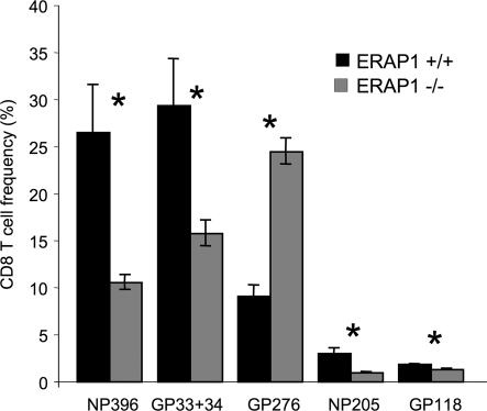
CD8(+) T cells respond to short peptides bound to MHC class I molecules. Although most antigenic proteins contain many sequences that could bind to MHC class I, few of these peptides actually stimulate CD8(+) T cell responses. Moreover, the T cell responses that are generated often follow a very reproducible hierarchy to different peptides for reasons that are poorly understood. We find that the loss of a single enzyme, endoplasmic reticulum aminopeptidase 1 (ERAP1), in the antigen-processing pathway results in a marked shift in the hierarchy of immunodominance in viral infections, even when the responding T cells have the same T cell receptor repertoire. In mice, ERAP1 is the major enzyme that trims precursor peptides in the endoplasmic reticulum and, in this process, can generate or destroy antigenic peptides. Consequently, when ERAP1 is lost, the immune response to some viral peptides is reduced, to others increased, and to yet others unchanged. Therefore, many epitopes must be initially generated as precursors that are normally trimmed by ERAP1 before binding to MHC class I, whereas others are normally degraded by ERAP1 to lengths that are too short to bind to MHC class I. Moreover, peptide trimming and the resulting abundance of peptide-MHC complexes are dominant factors in establishing immunodominance.


Conflict of interest statement

Conflict of interest statement: No conflicts declared.

Figures

Fig. 1.
Fig. 1.
ERAP1−/− mouse embryonic fibroblast lines are defective for peptide trimming in the ER. Fibroblast cell lines were prepared from ERAP1+/+ (black bars) and ERAP1−/− (gray bars) embryonic mice. (A) Cells were stained with mAb directed against H-2Kb (mAb B8.24.3) or H-2Db (28.14.8S) and analyzed by flow cytometry. (B) MEF cells were transfected with plasmids expressing SIINFEKL as a cytosolic ubiquitin fusion (S8L), ovalbumin protein targeted to the cytosol (Ova), LEQLE-SIINFEKL as a cytosolic ubiquitin fusion (N5-S8L), or ALEQLE-SIINFEKL targeted to the ER by a signal sequence (ss.N5-S8L). After 48 h, the cells were stained with mAb 25.D1.16 (anti-H-2Kb-SIINFEKL) and analyzed by flow cytometry. Data (representative of five experiments) show the mean fluorescence intensity with background staining by an irrelevant antibody subtracted (averages of three independent cell lines; error bars represent 1 SD). Asterisks indicate statistically significant differences (P < 0.05, Student t test) between ERAP1+/+ and ERAP1−/− MEF cell lines.
Fig. 2.
Fig. 2.
Splenocytes from ERAP1−/− mice express lower cell-surface MHC class I than those from ERAP1+/+ mice. Splenocytes from ERAP1+/+ mice (WT, black bars) or ERAP1−/− mice (KO, gray bars) were stained with mAb to H-2Kb or H-2Db or, as a control, to I-Ab and various antibodies to distinguish splenocyte subsets (CD11c, predominantly dendritic cells; CD8+and CD4, predominantly T lymphocyte subsets; B220, predominantly B cells) as indicated on the x axis. Shown are mean fluorescence intensities with background staining by an irrelevant antibody subtracted (averages of three mice; error bars represent 1 SD; representative of five experiments). Asterisks indicate statistically significant differences (P < 0.05, Student t test) between ERAP1+/+ and ERAP1−/− splenocytes.
Fig. 3.
Fig. 3.
CD8+ T cell responses to vaccinia virus are altered in ERAP1−/− mice. ERAP1+/+ (black bars) and ERAP1−/− (gray bars) mice were infected with recombinant vaccinia virus expressing full-length ovalbumin (Vac-FLOva) (A) or SIINFEKL targeted to the ER by a signal sequence (Vac-ss-SIINFEKL) (B). Eight days later, splenocytes were stimulated with peptides corresponding to vaccinia epitopes (B8R and A47L) or ovalbumin epitopes (SIINFEKL and KVVRFDKL), permeabilized, stained with anti-IFNγ antibody, and analyzed by flow cytometry. Shown is the percentage of CD8+ T cells producing IFNγ in response to each peptide (averages of three mice; error bars represent 1 SD; representative of at least three experiments). Asterisks indicate statistically significant differences (P < 0.05, Student t test) between ERAP1+/+ and ERAP1−/− mice.
Fig. 4.
Fig. 4.
CD8+ T cell responses to LCMV are altered in ERAP1−/− mice. ERAP1+/+ (black bars) or ERAP1−/− (gray bars) mice were infected with LCMV. Eight days later, splenocytes were stimulated with peptides corresponding to LCMV epitopes, permeabilized, and stained with anti-IFNγ antibody (A) or stained with tetramers consisting of H-2Kb or H-2Db complexed with the indicated LCMV epitopes and analyzed by flow cytometry (B). Shown is the percentage of CD8+ T cells reactive with each peptide (averages of three mice; error bars represent 1 SD; representative of at least three experiments). Asterisks indicate statistically significant differences (P < 0.05, Student t test) between ERAP1+/+ and ERAP1−/− mice. (Inset) Same data as in A and B, arranged to show the normal and altered immunodominance hierarchies.
Fig. 5.
Fig. 5.
Altered CD8+ T cell responses in ERAP1−/− mice are not due to altered T cell repertoire. Splenocytes from LCMV-immune B6/SJL mice were adoptively transferred into ERAP1+/+ (black bars) or ERAP1−/− (gray bars) mice. One day after transfer, recipient mice were infected with LCMV, and the recall CD8+ T cell response generated by the donor cells was analyzed by intracellular IFNγ staining. Shown is the percentage of CD8+ CD45.1+ T cells producing IFNγ in response to each peptide (averages of three mice; error bars represent 1 SD; representative of two experiments). Asterisks indicate statistically significant differences (P < 0.05, Student t test) between ERAP1+/+ and ERAP1−/− mice.

References

    1. Yewdell J. W., Bennink J. R. Annu. Rev. Immunol. 1999;17:51–88. - PubMed
    1. Busch D. H., Pamer E. G. J. Immunol. 1998;160:4441–4448. - PubMed
    1. Sandberg J. K., Grufman P., Wolpert E. Z., Franksson L., Chambers B. J., Karre K. J. Immunol. 1998;160:3163–3169. - PubMed
    1. Brehm M. A., Pinto A. K., Daniels K. A., Schneck J. P., Welsh R. M., Selin L. K. Nat. Immunol. 2002;3:627–634. - PubMed
    1. Wherry E. J., Blattman J. N., Murali-Krishna K., van der Most R., Ahmed R. J. Virol. 2003;77:4911–4927. - PMC - PubMed

Publication types

MeSH terms