. 2006 Jun 13;103(24):9232-7.
doi: 10.1073/pnas.0602956103. Epub 2006 Jun 5.

Estrogens protect pancreatic beta-cells from apoptosis and prevent insulin-deficient diabetes mellitus in mice

Affiliations

Estrogens protect pancreatic beta-cells from apoptosis and prevent insulin-deficient diabetes mellitus in mice

Cedric Le May et al. Proc Natl Acad Sci U S A. .

Abstract

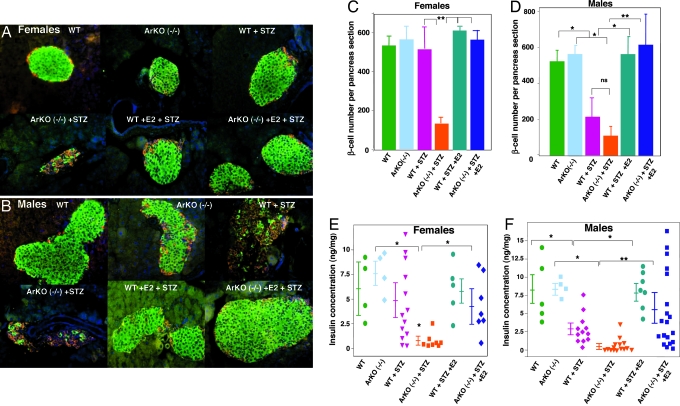
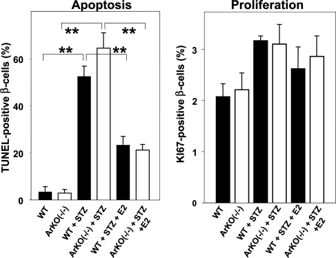
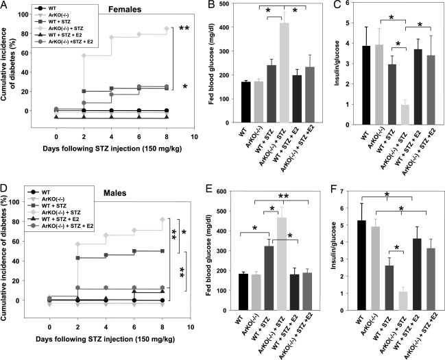
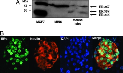
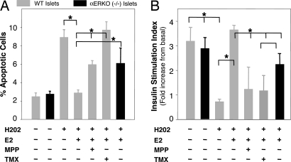
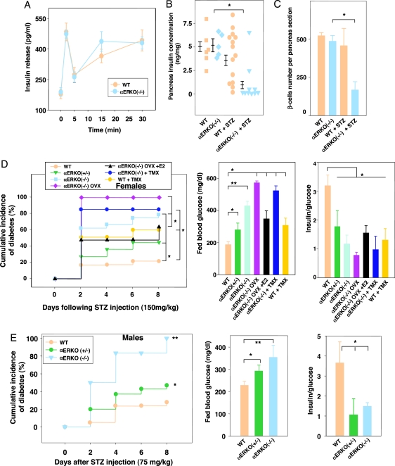
In diabetes, the death of insulin-producing beta-cells by apoptosis leads to insulin deficiency. The lower prevalence of diabetes in females suggests that female sex steroids protect from beta-cell injury. Consistent with this hypothesis, 17beta-estradiol (estradiol) manifests antidiabetic actions in humans and rodents. In addition, estradiol has antiapoptotic actions in cells that are mediated by the estrogen receptor-a (ERalpha), raising the prospect that estradiol antidiabetic function may be due, in part, to a protection of beta-cell apoptosis via ERalpha. To address this question, we have used mice that were rendered estradiol-deficient or estradiol-resistant by targeted disruption of aromatase (ArKO) or ERalpha (alphaERKO) respectively. We show here that in both genders, ArKO(-/-) mice are vulnerable to beta-cell apoptosis and prone to insulin-deficient diabetes after exposure to acute oxidative stress with streptozotocin. In these mice, estradiol treatment rescues streptozotocin-induced beta-cell apoptosis, helps sustain insulin production, and prevents diabetes. In vitro, in mouse pancreatic islets and beta-cells exposed to oxidative stress, estradiol prevents apoptosis and protects insulin secretion. Estradiol protection is partially lost in beta-cells and islets treated with an ERalpha antagonist and in alphaERKO islets. Accordingly, alphaERKO mice are no longer protected by estradiol and display a gender nonspecific susceptibility to oxidative injury, precipitating beta-cell apoptosis and insulin-deficient diabetes. Finally, the predisposition to insulin deficiency can be mimicked in WT mice by pharmacological inhibition of ERalpha by using the antagonist tamoxifen. This study demonstrates that estradiol, acting, at least in part, through ERalpha, protects beta-cells from oxidative injury and prevents diabetes in mice of both genders.


Conflict of interest statement

Conflict of interest statement: No conflicts declared.

Figures

Fig. 1.
Fig. 1.
Disruption of islet architecture and function in ArKO−/− mice exposed to STZ. (A and B) Representative pancreatic sections showing immunofluorescent staining for insulin (green) and glucagon (red) was performed in the indicated mice in basal conditions (vehicle) and after STZ injection (day 8). (C and D) β-cell number per pancreas section (n = 4–5) was calculated from pancreatic sections from A and B. (E and F) Pancreas insulin concentration was measured as described (24) in basal conditions and after STZ challenge (day 8). Values are represented as scatter plot and mean ± SE in ng/mg of pancreas (n = 4–19). ∗, P < 0.05; ∗∗, P < 0.01.
Fig. 2.
Fig. 2.
Estradiol protects mice from STZ-induced β-cell apoptosis in vivo. Apoptosis (Left) and proliferation (Right) were determined by TUNEL and KI67 immunostaining from pancreas section after vehicle or STZ injection in male mice. ∗∗, P < 0.01.
Fig. 3.
Fig. 3.
ArKO−/− mice are predisposed to STZ-induced insulin-deficient diabetes. Cumulative incidence of diabetes (random-fed blood glucose >250 mg/dl) was calculated by Kaplan–Meier estimation in female (A) (n = 10–28) and male (D) (n = 10–30) mice after STZ challenge (150 mg/kg of body weight). (B and E) Random-fed blood glucose (day 8), in female (B) and male (E) mice. (C and F) The ratio of random-fed of insulin (pg/ml) and glucose (mg/dl) at day 8 was used as an index of insulin deficiency in female (C) and male (F) mice. ∗, P < 0.05; ∗∗, P < 0.01.
Fig. 4.
Fig. 4.
ERα is expressed in β-cells. (A) Western blot analysis of ERα expression in MCF7 cells, MIN6, and mouse islets. (B) Female pancreas section showing a single islet with ERα nuclear staining in β-cells (ERα). The insulin (red), nuclear (blue) (DAPI), and triple staining (merge) are shown.
Fig. 5.
Fig. 5.
ERα is important for β-cell survival and function after exposure to oxidative stress. (A) WT or αERKO mouse islets were incubated with E2 (10−8 M), MPP (10−7 M), and TMX (10−7 M) for 72 h, followed by H2O2 (100 μM) for the last 6 h. Percent of apoptotic cells was assessed by flow cytometry by using TUNEL/PI double labeling. Values represent five independent experiments. (B) Insulin secretion in response to glucose was assessed in static incubations from WT or αERKO−/− mouse islets cultured with E2 (10−8 M), MPP (10−7 M), and TMX (10−7 M) for 72 h, followed by H2O2 (100 μM) for the last hour. The stimulation index represents the ratio of insulin released at 16.7 mM glucose to insulin released at 2.8 mM glucose in four to seven replicate experiments). ∗, P < 0.05.
Fig. 6.
Fig. 6.
αERKO mice are predisposed to STZ-induced insulin-deficient diabetes. (A) Acute-phase insulin secretion after i.p. glucose injection (3 g/kg of body weight) in female mice (n = 8–10) was measured as described (24). (B) Pancreas insulin concentration was measured in female mice at basal levels or after 150 mg/kg STZ injection (day 8). Values are represented as scatter plot and mean ± SE (n = 5–14). (C) β-cell number per pancreas section at basal levels and after STZ injection in female mice at day 8 (n = 4). (D and E) (Left) Cumulative incidence of diabetes in mice after STZ injection. (Center) Random-fed blood glucose in mice (day 8). (Right) Ratio of random-fed insulin (pg/ml) to glucose (mg/dl) in female and male mice after STZ injection (day 8). ∗, P < 0.05; ∗∗, P < 0.01.

References

    1. Mathis D., Vence L., Benoist C. Nature. 2001;414:792–798. - PubMed
    1. Robertson R. P., Harmon J., Tran P. O., Tanaka Y., Takahashi H. Diabetes. 2003;52:581–587. - PubMed
    1. Wild S., Roglic G., Green A., Sicree R., King H. Diabetes Care. 2004;27:1047–1053. - PubMed
    1. Gale E. A., Gillespie K. M. Diabetologia. 2001;44:3–15. - PubMed
    1. Mauvais-Jarvis F., Sobngwi E., Porcher R., Riveline J. P., Kevorkian J. P., Vaisse C., Charpentier G., Guillausseau P. J., Vexiau P., Gautier J. F. Diabetes. 2004;53:645–653. - PubMed

Publication types

MeSH terms