. 2006 Jun 13;103(24):9034-8.
doi: 10.1073/pnas.0602859103. Epub 2006 Jun 5.

Centrosome-associated RNA in surf clam oocytes

Affiliations

Centrosome-associated RNA in surf clam oocytes

Mark C Alliegro et al. Proc Natl Acad Sci U S A. .

Abstract

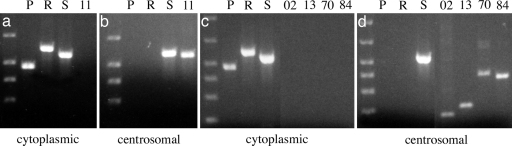
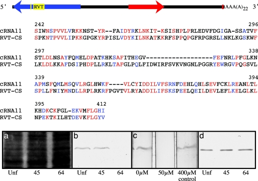
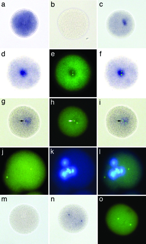
Centrosomes are the major microtubule-organizing center in animal cells. They are composed of a pair of [9(3) + 0] centrioles surrounded by a relatively ill-defined pericentriolar matrix, provide the ciliary centriole-kinetosome (basal body) progenitor, and organize the assembly of microtubules into the mitotic spindle during cell division. Despite >100 years of microscopic observation and their obvious significance, our understanding of centrosome composition, dynamic organization, and mechanism of action is limited when compared with that of other cellular organelles. Centrosomes duplicate only once per cell cycle to ensure development of a normal bipolar spindle. The initial event in centrosome duplication is centriole replication, which is generative, semiconservative, and independent of the nucleus. Such observations led to the proposal that centrosomes contain their own complement of nucleic acids, possibly representative of an organellar genome comparable with those described for mitochondria and chloroplasts. The consensus in the field is that centrosomes lack DNA but may contain RNA. We isolated centrosomes from oocytes of the surf clam, Spisula solidissima, and purified from them a unique set of RNAs. We show here by biochemical means and subcellular in situ hybridization that the first transcript we analyzed is intimately associated with centrosomes. Sequence analysis reveals that this centrosome-associated RNA encodes a conserved RNA-directed polymerase domain. The hypothesis that centrosomes contain an intrinsic complement of specific RNAs suggests new opportunities to address the century-old problem of centrosome function, heredity, and evolution.


Conflict of interest statement

Conflict of interest statement: No conflicts declared.

Figures

Fig. 1.
Fig. 1.
Differential expression of cnRNAs in centrosomes vs. whole oocyte cytoplasm. Equal quantities of RNA isolated from either whole oocyte cytoplasm or centrosomal fractions were reverse transcribed and used as template in PCR with primers directed against known cytoplasmic RNAs or putative cnRNAs. Conditions such as number of cycles, template input, and product sizes were chosen empirically to yield product in the linear range of amplification. The results of two separate experiments are shown. (a and b) The distribution of cnRNA11 between cytoplasmic and centrosomal fractions, respectively. (c and d) The distribution between cytoplasmic and centrosomal fractions of cnRNAs 102 (02), 113 (13), 170 (70), and 184 (84). The first lane in each panel is a 100-bp DNA reference ladder. P, poly(A)-binding protein RNA; R, ribonucleotide reductase; S, 18s rRNA subunit. Trace quantities of cnRNA PCR product can sometimes be seen in cytoplasmic template lanes but are not visible in these panels.
Fig. 2.
Fig. 2.
Structure of cnRNA11. (Top) A canonical polyadenylation signal 29 nucleotides upstream from the poly(A) tail is represented by the small red box near the 3′ end in the diagrammatic representation shown. The red arrow represents the 638-nt sequence originally cloned from centrosome preparations, 5′ to 3′ orientation (this region was used to generate probe for the in situ hybridizations shown in Fig. 3). An ORF predicting a 54-kDa polypeptide discovered in the antisense strand (arrowhead indicating direction from 3′ to 5′ relative to the sense strand) is shown in blue, with RNA-dependent polymerase (RVT) and RNP-1 domains shown in yellow. (Middle) Alignment of the cnRNA11 reverse transcriptase domain with the pfam00078 reverse transcriptase consensus sequence. Identities are shown in red, and conservative substitutions are shown in blue. Bit score = 94.7, P = 5e−21, GenBank accession no. DQ359732. (Bottom) Western blot analysis of cnRNA11 protein expression in early embryos. (a) A segment of Coomassie blue-stained polyacrylamide gel indicating relative protein loads (50 μg per lane) for lysed unfertilized (Unf) oocytes and 45 and 64 min zygotes. (b) A blot showing relative levels of cnRNA11 protein at these three developmental time points. Controls for specificity included preimmune serum as well as preabsorption of immune serum with the antigenic peptide. Results with preimmune serum were negative (not shown). Preabsorption controls are shown in c, which depicts antibody staining of three blot strips in the absence of peptide (0 μM), after incubation of serum with 50 μM antigenic peptide to preabsorb immunoreactivity, and after incubation with 400 μM nonrelevant peptide, which had no effect on immunoreactivity. (d) A Western blot using an antibody to an unknown Spisula protein and, in addition to a, serves as a loading control for the down-regulation of cnRNA11 protein depicted in b.
Fig. 3.
Fig. 3.
In situ localization of cnRNA11. Spisula zygote labeled with antisense (a) and sense (b) probes for poly(A)-binding protein. Intense stain for poly(A)-binding protein mRNA is widely distributed in the cytoplasm. (c) Zygote hybridized with cnRNA11 probe complementary to the ORF shows single patch labeling pattern. (df) Another zygote labeled with this cnRNA11 hybridization probe (d) and colabeled with antibodies to the centrosome marker protein, γ-tubulin (e). An overlay of d and e is shown in f. The two γ-tubulin-labeled centrosomes are encased within the cnRNA11 hybridization patch. The cell depicted in gi is from a sample developed for only 3 days as opposed to the typical 6-day development period for our in situ hybridizations. A densely stained cnRNA11 puncta is clearly visible (arrow in g), which coincides precisely with the γ-tubulin-labeled centrosome seen in h; an overlay of g and h is shown in i. The hybridization protocol used for labeling the cells (ai) included 1% SDS in the prehybridization and hybridization steps. Chromatin structure and protein antigenicity are significantly compromised in these samples, although the monoclonal anti-γ-tubulin antibody used here was effective. (jl) An embryo labeled (green) by fluorescent step-down in situ hybridization. Protein antigenicity is destroyed in the step-down method, but chromatin organization, although impaired, is better preserved than in SDS-treated embryos. A mitotic figure is discernable in the Hoechst-stained image of this cell (k). (l) Overlay of j and k. (m) Example of a zygote hybridized with cnRNA11 control probe; hybridization signal was not seen at any stage after activation. (n) Zygote fixed 16 min after activation and labeled with hybridization probe complementary to the cnRNA11 ORF by using the SDS protocol. Two distinct “kernel” structures are visible. These kernels did not stain with anti-γ-tubulin. Another 16-min zygote, prepared for routine immunocytochemistry and labeled with anti-γ-tubulin, is shown in o to compare the appearance and spatial organization of centrosomes with cnRNA11 kernels at the same time point. A nonspecific background signal is seen in all fluorescent images, attributable to the paraformaldehyde-fixed chorion.

References

    1. Sluder G., Hinchcliffe E. H. Biol. Cell. 1999;91:413–427. - PubMed
    1. Wong C., Stearns T. Nat. Cell Biol. 2003;5:539–544. - PubMed
    1. Chapman M. J., Dolan M. F., Margulis L. Q. Rev. Biol. 2000;75:409–429. - PubMed
    1. Stich H. Chromosoma. 1954;6:199–236. - PubMed
    1. Mazia D. Symp. Soc. Exp. Biol. 1955;9:335–357.

Publication types