. 2006 Jun 13;103(24):9250-5.
doi: 10.1073/pnas.0602473103. Epub 2006 Jun 5.

Smad3 deficiency alters key structural elements of the extracellular matrix and mechanotransduction of wound closure

Affiliations

Smad3 deficiency alters key structural elements of the extracellular matrix and mechanotransduction of wound closure

Praveen R Arany et al. Proc Natl Acad Sci U S A. .

Abstract

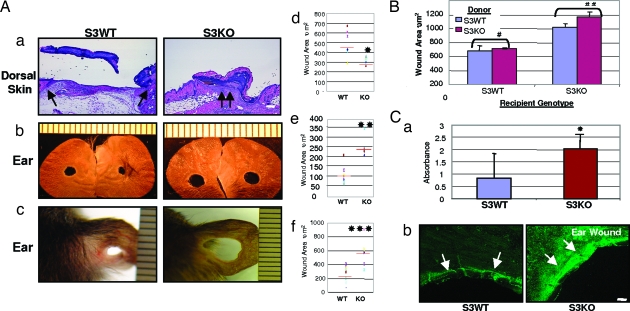
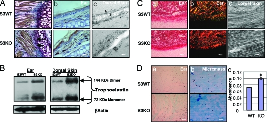
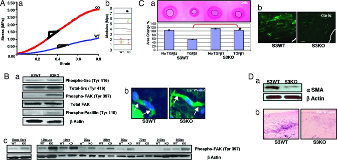
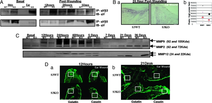
The loss of TGFbeta or its downstream mediator, Smad3, key players in tissue repair, accelerates closure of incisional wounds in mice. In contrast, we now report that excisional ear wounds in mice lacking Smad3 enlarge compared with wild-type controls resulting from changes in extracellular matrix molecules, which alter the mechanotransduction properties of these wounds. Specifically, levels of elastin and glycosoaminoglycans are increased, collagen fibers are more compactly organized, and matrix modulators like integrins, TGFbeta1, and matrix metalloproteinases (MMPs) are altered both basally and after wounding in Smad3 knockout mice. Mechanical testing of dorsal skin correlates these changes in matrix composition with functional parameters, specifically an increased elastic modulus, suggesting an imbalance of tissue forces. We propose that the altered mechanical elastic properties translate into a persistent retractile force that is opposed by decreased wound contractile forces contributing to the enlarging ear wound in Smad3 knockout mice. These studies highlight a previously undescribed role for Smad3 in the mechanotransduction of matrix unsupported ear wound closure.


Conflict of interest statement

Conflict of interest statement: No conflicts declared.

Figures

Fig. 1.
Fig. 1.
Excisional would healing in Smad3 mice. (A) Smad3-null mice demonstrate opposite wound healing responses in the dorsal skin vs. the ear. (a) Dorsal skin 48 h after wounding stained with hematoxylin/eosin. Arrows point to epithelial wound margins. (Scale bar: 100 μm.) (b) Ear wounds 38 days after wounding. (c) Ear wounds in SVEV129 mice at 36 days after wounding. (df) Quantification of wound area using a scatter plot. Red bar represents means; n = 6–8 mice, ∗, P < 0.05; n = 3–4, ∗∗, P < 0.0001; and ∗∗∗, P < 0.05. (B) Quantification of wound area after bone marrow transplantation between genotypes in the healing ear wound at 21 days. n = 3 mice. Shown are means ± SD. Healing differences after homologous marrow transplants were not statistically significant: #, P > 0.5, and ##, P > 0.1. (C) (a) Quantification of apoptotic cell death in ear wounds by ELISA data represented as means ± SD. n = 5; ∗, P < 0.05. (b) TUNEL staining of ear wounds at 24 h. Arrows point to a subepithelial prominent band of dermal apoptotic cell at wound edge. (Scale bar: 100 μm.)
Fig. 2.
Fig. 2.
Matrix components are basally perturbed in S3KO mice. (A) Elastin is increased in S3KO tissues as demonstrated by the following. (a) Orcein Picroindigocaramine staining. Arrows point to brown coarse elastin fibers tethering collagen fibers to perichondrium in ear. (b) Immunohistochemistry in ear. (Scale bars for a and b: 25 μm.) (c) EM of dorsal skin demonstrates increased tropoelastin (T) core and disorganized microfibrillar (M) peripheral sheath of elastin fibers in S3KO dorsal skin. (Scale bar: 0.2 μm.) (B) Western blot for elastin. (C) Collagen fibers stained by Picrosirius staining and polarizing microscopy. (a) Phase contrast. (b) Polarizing filters. (Scale bars for a and b: 50 μm.) (c) EM of dorsal skin revealed condensed fibrillar arrangement. (Scale bar: 0.2 μm.) (D) (a) GAGs visualized by alcian blue (pH 1) staining in ears. (Scale bar: 125 μm.) (b) Micromass cultures of primary DFs. (Scale bar: 50 μm.) (c) Quantification of GAG expression in these cells by spectrophotometery, showing means ± SD. ∗, P < 0.01.
Fig. 3.
Fig. 3.
Tissue mechanical properties and forces are altered in S3KO mice. (A) (a) Stress vs. strain plots of dorsal skin in linear region in representative sample curves. (b) Scatterplot of modulus of individual samples. Red bar represents means; n = 7 or 8 samples; ∗, P < 0.05. (B) (a) Wound tissue analyzed for stress-associated signaling and adhesion molecules at 12 h after wounding. (b) Stress fibers at 24 h after wounding in ear tissues demonstrated (arrows) in individual fibroblasts by phalloidin staining and counterstained with DAPI. (Scale bar: 7 μm.) (c) Time course analysis of phospho-FAK levels by Western blots. (C) (a) Primary DFs in annular collagen–elastin gels in the presence or absence of TGFβ1 (2.5 ng/ml) at 5 days (Upper) and quantification of contracted central annular gel area (Lower). Shown are means ± SD; n = 3; ∗, P < 0.001. (b) αSMA immunostaining in gels. (Scale bar: 100 μm.) (D) Wound tissues at 7 days analyzed for αSMA by Western blotting (a) and immunostaining (b). (Scale bar: 125 μm.)
Fig. 4.
Fig. 4.
Matrix modulators are perturbed in S3KO. (A) Immunoprecipitations with tissue extracts performed with specific integrin complex antibodies, αVβ3 and αVβ5, and immunoblotted with αV antibody. (B) Wound margins analyzed for TGFβ1 at 24 h by immunostaining (a) and quantification (b). Red bar represents means; n = 5; ∗, P < 0.05. (Scale bar: 125 μm.) (C) Time course substrate-specific zymography for MMP2 and MMP9 (gelatin; Upper) or MMP12 (casein; Lower). (D) In situ zymography on cryosections using fluorescent substrate overlays 12 h (a) and 21 days (b) after wounding. (Scale bars: 200 μm.) Boxed areas show prominent enzymatic activity.

References

    1. Kim L. T., Yamada K. M. Proc. Soc. Exp. Biol. Med. 1997;214:123–131. - PubMed
    1. Koch R. M., Roche N. S., Parks W. T., Ashcroft G. S., Letterio J. J., Roberts A. B. Wound Repair Regen. 2000;8:179–191. - PubMed
    1. Ashcroft G. S., Yang X., Glick A. B., Weinstein M., Letterio J. L., Mizel D. E., Anzano M., Greenwell-Wild T., Wahl S. M., Deng C., et al. Nat. Cell Biol. 1999;1:260–266. - PubMed
    1. Falanga V., Schrayer D., Cha J., Butmarc J., Carson P., Roberts A. B., Kim S. J. Wound Repair Regen. 2004;12:320–326. - PubMed
    1. Clark L. D., Clark R. K., Heber-Katz E. Clin. Immunol. Immunopathol. 1998;88:35–45. - PubMed

Publication types

MeSH terms

LinkOut - more resources