. 2006 Jun 13;103(24):9017-22.
doi: 10.1073/pnas.0508825103. Epub 2006 Jun 5.

Semaphorin 4D provides a link between axon guidance processes and tumor-induced angiogenesis

Affiliations

Semaphorin 4D provides a link between axon guidance processes and tumor-induced angiogenesis

John R Basile et al. Proc Natl Acad Sci U S A. .

Abstract

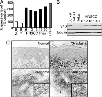
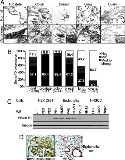
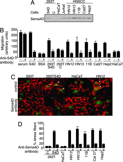
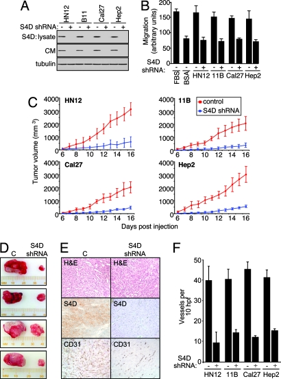
Tumor progression and metastasis depend on the ability of cancer cells to initiate angiogenesis and ensure delivery of oxygen, nutrients, and growth factors to rapidly dividing transformed cells and provide access to the systemic circulation. In addition to well established growth factors and inflammatory mediators that promote capillary sprouting and endothelial cell growth and migration, an emerging body of evidence supports a previously unrecognized function for axon guidance molecules in regulation of blood vessel development. Here we show that semaphorin 4D (Sema4D), a protein originally shown to regulate axonal growth cone guidance in the developing central nervous system through its receptor, plexin-B1, is highly expressed in cell lines derived from head and neck squamous cell carcinomas (HNSCCs) at both the protein and message level. Immunohistochemical analysis of a large collection of HNSCC specimens revealed high levels of Sema4D in a cell surface pattern in invading islands of transformed epithelial cells, but not in normal and noninvasive dysplastic epithelium. A similar pattern was observed in malignant cells from prostate, colon, breast, and lung cancer tissues. When shed from HNSCC cells, Sema4D stimulates endothelial cell migration, which can be prevented by Sema4D-blocking antibodies and by Sema4D knockdown. Furthermore, knocking down Sema4D by lentiviral expression of Sema4D shRNA reduces dramatically the size and vascularity of HNSCC tumor xenografts. These findings indicate that expression of Sema4D is a frequently used strategy by which a wide variety of carcinomas may promote angiogenesis, and therefore is a possible therapeutic target for the treatment of these malignancies.


Conflict of interest statement

Conflict of interest statement: No conflicts declared.

Figures

Fig. 1.
Fig. 1.
Sema4D is highly expressed in HNSCC. (A) RNAs from controls and HNSCC cell lines were compared to reference RNA by microarray analysis. Sema4D mRNA levels are expressed relative to normal human oral keratinocytes (NOK). RNA from brain tissues was used as the positive control. IOK, immortal human oral keratinocytes. (B) Immunoblot analysis for Sema4D shows a band at ≈125 kDa for endogenously expressed protein in lysates from Jurkat cells (positive control) and HNSCC cell lines, but not in the nontumorigenic epithelial-derived cell lines HaCaT or HeLa. Tubulin was used as a loading control. (C) Immunohistochemical analysis of Sema4D in HNSCC. No Sema4D was detected in normal parakeratinized stratified squamous epithelium (Normal) and in dysplastic stratified squamous epithelium, although chronic inflammatory cells were positive (Dysplastic). Sema4D immunoreactivity is seen in invading islands of transformed epithelial cells (Carcinoma). A cell surface-staining pattern is seen at a higher magnification (Insets).
Fig. 2.
Fig. 2.
Sema4D is expressed in oral, prostate, colon, breast, and lung cancer, whereas plexin-B1 is expressed in tumor endothelial cells. (A) Tumor tissue arrays were analyzed for Sema4D expression. The photographs show representative areas of the indicated tumors and corresponding normal tissues. Normal tissues (Upper) exhibit no epithelial Sema4D expression or small foci in areas of areas of chronic inflammatory cell accumulation (normal colon). Tumor tissues demonstrate abundant Sema4D staining in invading islands of transformed epithelial cells in prostate, colon, breast, and lung carcinomas, although not in ovarian carcinomas (Lower). A robust cell-surface-specific staining pattern for Sema4D is seen at higher magnification (Insets). (B) The bar graph summarizes the data from Sema4D staining in tumor tissue arrays. Between 37.5% and 85.4% of oral, prostate, colon, breast, and lung carcinomas analyzed exhibit moderate to strong Sema4D expression. (C) Immunoblot analysis for plexin-B1 in the indicated endothelial and HNSCC cell lines. (Upper) Control transfected HEK293T cells or cells transfected with full-length plexin-B3, -B2, and –B1 demonstrate antibody specificity. (Lower) Tubulin was used as a loading control. (D) Immunohistochemical analysis of plexin-B1 in tumor tissues. (Right) Abundant plexin-B1 staining is seen in the endothelial cells lining the blood vessels. (Left) A cross section of peripheral nerve is shown as a positive control.
Fig. 3.
Fig. 3.
Sema4D shed by HNSCC cells promotes stress fiber formation and chemotaxis in endothelial cells. (A) Immunoblot analysis for Sema4D in medium conditioned by the indicated cell lines and HEK293T cells transfected with Sema4D (S4D) or vector controls (C). Sema4D is seen in medium conditioned by the HNSCC cell lines but not in medium conditioned by HaCaT. Jurkat cells were used as a positive control. (B) Serum-free medium containing the indicated cell type or protein, with or without 10 μg/ml Sema4D-blocking antibody, were used as the chemoattractants for endothelial cells in a migration assay. Ten percent FBS and purified Sema4D were used as positive controls and 0.1% BSA was the negative control. HEK293T cells transfected with Sema4D (S4D) or vector (C) were used as additional controls. (C) HUVEC stably expressing EGFP were cocultured with HEK293T cells transfected with Sema4D (S4D) or vector (C), HaCaT, or HN12 cells with or without Sema4D-blocking antibody (C and Sema4D antibody, respectively), stained with Texas red-phalloidin and viewed in an immunofluorescence microscope. Endothelial cells exhibiting stress fibers are indicated with an arrowhead. (D) Quantification of stress fiber formation in EGFP-positive HUVEC cultured with the indicated cell lines and vector (C) or Sema4D (S4D) transfected HEK293T, with (+) and without (−) Sema4D-blocking antibody. The number of endothelial cells exhibiting stress fiber polymerization in the cocultures was counted and expressed as a percentage of the total endothelial cells in the cocultures.
Fig. 4.
Fig. 4.
Sema4D knockdown reduces HNSCC-mediated endothelial cell chemotaxis and HNSCC tumor growth and vascularity. (A) Immunoblot analysis for Sema4D on cell lysates (S4D: lysate) and serum-free medium (CM) conditioned by the indicated cell types transfected with vector control (−) or Sema4D shRNA (+) (Top and Middle). Tubulin was used as a lysate loading control (Bottom). (B) Serum-free media conditioned by control (−) or Sema4D shRNA (+) transfected HNSCC cells were used as the chemoattractants for HUVEC in migration assays. Ten percent FBS and 0.1% BSA served as positive and negative controls. (C) The HNSCC cells HN12, UMSCC 11B, Cal27, and Hep2 were infected with control lentiviruses or lentiviruses coding for Sema4D shRNA and injected s.c. into nude mice. The results of tumor volume measurement from each day of the experiment are shown (n = 10). (D) Representative tumors derived from HN12 cells infected with EGFP or Sema4D lentiviruses are shown at the time of killing. (E) Tumors derived from HN12 cells infected with a control lentivirus (C, Left) and tumors from Sema4D shRNA-infected cells (S4D shRNA, Right) are shown in hematoxylin- and eosin-stained sections (H&E). Enhanced blood vessel infiltration is seen in tumors derived from control HN12 cells compared with tumors derived from Sema4D shRNA-infected cells (CD31). Tumors of HN12 cells infected with control lentiviruses also exhibit much higher levels of Sema4D staining compared with Sema4D shRNA-infected cells (S4D). (F) The number of vessels per 10 high-power fields in CD31-stained xenografts derived from Sema4D shRNA- and control-infected HNSCC cells was counted and averaged.

References

    1. Bergers G., Benjamin L. E. Nat. Rev. Cancer. 2003;3:401–410. - PubMed
    1. Carmeliet P. Nat. Med. 2000;6:389–395. - PubMed
    1. Folkman J. Nat. Med. 1995;1:27–31. - PubMed
    1. Auguste P., Gursel D. B., Lemiere S., Reimers D., Cuevas P., Carceller F., Di Santo J. P., Bikfalvi A. Cancer Res. 2001;61:1717–1726. - PubMed
    1. Dor Y., Porat R., Keshet E. Am. J. Physiol. 2001;280:C1367–C1374. - PubMed

MeSH terms