. 2006 Jun 13;103(24):9069-74.
doi: 10.1073/pnas.0603453103. Epub 2006 Jun 5.

Mutation of the atrophin2 gene in the zebrafish disrupts signaling by fibroblast growth factor during development of the inner ear

Affiliations

Mutation of the atrophin2 gene in the zebrafish disrupts signaling by fibroblast growth factor during development of the inner ear

Yukako Asai et al. Proc Natl Acad Sci U S A. .

Abstract

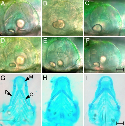
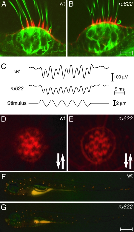
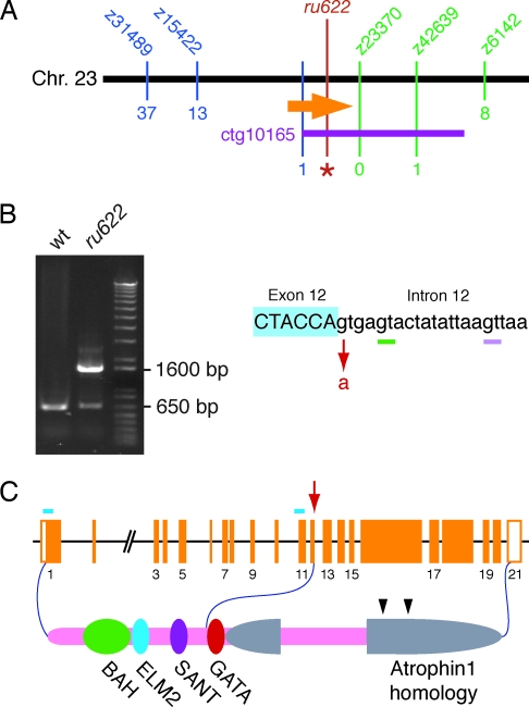
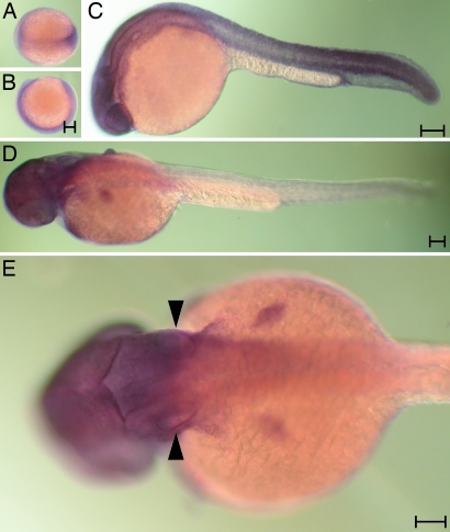
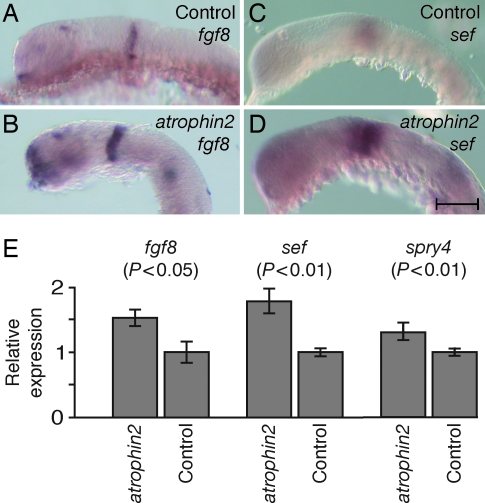
The development of the vertebrate inner ear depends on the precise expression of fibroblast growth factors. In a mutagenesis screen for zebrafish with abnormalities of inner-ear development and behavior, we isolated a mutant line, ru622, whose phenotypic characteristics resembled those of null mutants for the gene encoding fibroblast growth factor 8 (Fgf8): an inconsistent startle response, circular swimming, fused otoliths, and abnormal semicircular canals. Positional cloning disclosed that the mutant gene encodes the transcriptional corepressor Atrophin2. Both the Fgf8 protein and zebrafish "similar expression to fgf genes" protein (Sef), an antagonist of fibroblast growth factors induced by Fgf8 itself, were found to be overexpressed in ru622 mutants. We therefore hypothesized that an excess of Sef eliminates Fgf8 signals and produces an fgf8 null phenotype in ru622 mutants. In support of this idea, we could rescue larvae whose atrophin2 expression had been diminished with morpholinos by reducing the expression of Sef as well. We propose that Atrophin2 plays a role in the feedback regulation of Fgf8 signaling. When mutation of the atrophin2 gene results in the overexpression of both Fgf8 and Sef, the excessive Sef inhibits Fgf8 signaling. The resultant imbalance of Fgf8 and Sef signals then underlies the abnormal aural development observed in ru622.


Conflict of interest statement

Conflict of interest statement: No conflicts declared.

Figures

Fig. 1.
Fig. 1.
Morphological features of WT (A, D, and G), ru622 mutant (B, E, and H), and fgf8 knockdown (C, F, and I) larvae at 5 dpf. (A) The otic vesicle of a WT animal contains two otoliths, a smaller, anterior one lying in the horizontal plane and a larger, posterior one oriented vertically. (B and C) By contrast, an ru622 mutant (B) or a larva treated with a morpholino against fgf8 (C) sometimes has only a single otolith. (D) The semicircular canals of a WT animal begin to be delineated at this stage by three pillars extending from the anterior, posterior, and ventral walls of the otic vesicle and by a dorsolateral septum protruding from the dorsal wall to the center of the otic vesicle. (E and F) In an ru622 mutant (E) or an fgf8 knockdown larva (F), the formation of semicircular canals is abnormal. (G) Alcian blue staining reveals the pattern of cartilage formation in the rostral region of a WT larva. P, palatoquadrate cartilage; C, ceratohyal cartilage; M, Meckel's cartilage. (H and I) An ru622 mutant (H) or an fgf8 knockdown larva (I) is distinguished by shorter palatoquadrate cartilages and the orientation of the ceratohyal cartilages at an increased angle. (Scale bars: 20 μm for AF; 100 μm for GI.)
Fig. 2.
Fig. 2.
Phenotypic characteristics of the ru622 mutation. (A) In a confocal image of the crista in the lateral semicircular canal of a larva at 5 dpf, Alexa Fluor 568-phalloidin (red) labels the stereocilia and an antiserum against acetylated tubulin (green) labels the kinocilia and cellular cytoskeletons. (B) The crista of a mutant larva displays similar features. (C) Extracellular microphonic recordings at 5 dpf indicate that the transduction current of a WT otic vesicle is approximately twice that of an ru622 mutant. (D) In a confocal image of the apical surface of a WT neuromast stained with Alexa Fluor 568-phalloidin, the axis of each hair bundle's polarity is denoted by a notch in the actin-rich cuticular plate corresponding to the position of the kinocilium. The arrows indicate the polarities observed. (E) The hair bundles of an ru622 mutant display normal polarization. (D) Fluorescence imaging of a WT larva exposed to 4-(4-(diethylamino)styryl)-N-methylpyridinium iodide reveals a stereotyped pattern of labeled neuromasts. (F) The labeling pattern is similar in an ru622 mutant, but the average number of labeled neuromasts is smaller than in a WT larva. (Scale bars: 10 μm for A, B, D, and E; 500 μm for F and G.)
Fig. 3.
Fig. 3.
Identification of the basis of the ru622 mutation. (A) A map of the genomic region surrounding the ru622 locus on zebrafish chromosome 23 displays the number of recombinations with respect to five simple sequence-length polymorphisms (blue and green) and a single-nucleotide polymorphism near the end of the bacterial artificial chromosome contig ctg10165 (purple). A total of 2,247 larvae were examined for each marker save z6142, for which only 1,164 animals were analyzed. The orange arrow represents the atrophin2 gene and the red line and asterisk denote the ru622 mutation. (B) When WT RNA is used as a template, RT-PCR amplification of a cDNA region extending from exon 8 to exon 13 produces a product ≈650 bp in length (Left). A reaction using cDNA from ru622 mutants yields a product of similar size and another ≈1,600 bp long. The ru622 mutation occurred at the boundary between exon 12 and intron 12, where the splice donor GT was changed to AT (Right). The smaller RT-PCR band from the ru622 reaction includes two products. In one, splicing occurs 4 bp downstream from the mutated splice site (green bar). In the other, splicing instead occurs 16 bp downstream (pink bar). The large RT-PCR product contains the entirety of intron 12. (C) The genomic structure of the zebrafish atrophin2 gene is represented as orange boxes. The arrow indicates the site of the ru622 mutation; the blue bars denote the targets of morpholinos directed against the translation start site and the splicing site between intron 10 and exon 11. Atrophin2 includes a bromo-adjacent homology domain (BAH), an EGL27 and Mta1 homology 2 domain (ELM2), a SWI3, ADA2, N-CoR and TFIIIB domain (SANT), and a zinc finger domain (GATA). The two arrowheads indicate RE repeats.
Fig. 4.
Fig. 4.
The expression pattern of the atrophin2 gene. Whole-mount in situ hybridization was conducted with a 3′ probe at various developmental stages. (A and B) Labeling of the germ band is apparent in a dorsal (A) or a lateral view (B) of the late gastrula. (C) At 24 hpf, essentially all tissue except the yolk is labeled. (D) Labeling diminishes from the caudal extreme by 36 hpf. (E) Arrowheads highlight the labeling of the otic vesicles in a dorsal view of a larva at 36 hpf. (Scale bar: 100 μm.)
Fig. 5.
Fig. 5.
The expression of fgf8 and sef genes in WT and atrophin2 knockdown embryos. (A) In a control embryo at 24 hpf, fgf8 transcripts are detected in the telencephalon, optic stalk, isthmus, otic vesicle, developing somites, and tail bud. (B) fgf8 is expressed more broadly in an atrophin2 knockdown embryo. (C) A 26-somite control embryo treated with sef probe displays prominent labeling of the optic cup and otic vesicle. (D) An atrophin2 knockdown embryo shows a similar pattern of labeling but more extensive expression than in control fish. (E) Quantitative PCR analysis confirms the increased expression of messages for fgf8, sef, and sprouty4 in atrophin2 knockdown animals; significance values were obtained with Student's one-tailed t test. Control animals were injected with water. The error bars indicate standard deviations. (Scale bar: 100 μm.)

References

    1. Hudspeth A. J. Nature. 1998;341:397–404. - PubMed
    1. Barald K. F., Kelley M. W. Development (Cambridge, U.K.) 2004;131:4119–4130. - PubMed
    1. Haddon C., Lewis J. J. Comp. Neurol. 1996;365:113–128. - PubMed
    1. Whitfield T. T., Riley B. B., Chiang M.-Y., Phillips B. Dev. Dyn. 2002;223:427–458. - PubMed
    1. Haddon C., Lewis J. Development (Cambridge, U.K.) 1991;112:541–550. - PubMed

Publication types

MeSH terms

LinkOut - more resources