. 2006 Jun 5;173(5):651-8.
doi: 10.1083/jcb.200603092.

Ups1p, a conserved intermembrane space protein, regulates mitochondrial shape and alternative topogenesis of Mgm1p

Affiliations

Ups1p, a conserved intermembrane space protein, regulates mitochondrial shape and alternative topogenesis of Mgm1p

Hiromi Sesaki et al. J Cell Biol. .

Abstract

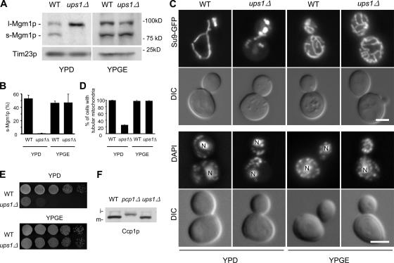
Mgm1p is a conserved dynamin-related GTPase required for fusion, morphology, inheritance, and the genome maintenance of mitochondria in Saccharomyces cerevisiae. Mgm1p undergoes unconventional processing to produce two functional isoforms by alternative topogenesis. Alternative topogenesis involves bifurcate sorting in the inner membrane and intramembrane proteolysis by the rhomboid protease Pcp1p. Here, we identify Ups1p, a novel mitochondrial protein required for the unique processing of Mgm1p and for normal mitochondrial shape. Our results demonstrate that Ups1p regulates the sorting of Mgm1p in the inner membrane. Consistent with its function, Ups1p is peripherally associated with the inner membrane in the intermembrane space. Moreover, the human homologue of Ups1p, PRELI, can fully replace Ups1p in yeast cells. Together, our findings provide a conserved mechanism for the alternative topogenesis of Mgm1p and control of mitochondrial morphology.


Figures

Figure 1.
Figure 1.
Identification of UPS1. (A) A model for alternative topogenesis of Mgm1p at the IM. The TIM23 translocon (gray), TM1 (blue), and TM2 (yellow) of Mgm1p, the mitochondrial processing peptidase (MPP, green) and Pcp1 protease (green) are shown. (B) WT cells were labeled with [35S]-methionine and -cysteine, and chased for the indicated time period. Proteins were examined by immunoprecipitation using anti-Mgm1p antibodies. (C) Cell lysates from WT and indicated mutant strains grown in YPGalSuc were analyzed by immunoblotting using anti-Mgm1p antibodies. (D) Isolated mitochondria were analyzed by immunoblotting using the indicated antibodies.
Figure 2.
Figure 2.
Phenotypes of ups1Δ cells. (A) Cell lysates from WT and ups1Δ cells grown in YPD or YPGE were analyzed by immnoblotting using antibodies to Mgm1p and Tim23p. (B) Mgm1p signals were quantitated by densitometry using NIH image. The percentage of s-Mgm1p was calculated from the ratio of s-Mgmp1/(s-Mgm1p + l-Mgm1p). Values are mean ± SD (n = 3). (C, top panel) Cells expressing mitochondria-targeted Su9-GFP (Westermann and Neupert, 2000) were grown in YPD or YPGE to log phase and viewed by differential interference contrast and fluorescence microscopy. (C, bottom panel) Cells were fixed in 3.7% glutaraldehyde, washed, and stained with 1 μg/ml DAPI. ‘N’ indicates nuclear DNA staining. Bars, 3 μm. (D) Quantitation of mitochondrial morphology (n = 300). (E) Serial dilutions of WT and ups1Δ cells were spotted onto YPD and YPGE and incubated at 30°C for 2 d and 4 d, respectively. (F) Lysates from cells expressing Ccp1p-HA grown in YPGalSuc were analyzed by immnoblotting using anti-HA antibodies. i: i-Ccp1p, m: m-Ccp1p.
Figure 3.
Figure 3.
The expression of PRELI rescues ups1Δ cells. (A) Phylogenetic analysis of Ups1p homologues in S. cerevisiae (Sc), S. pombe (Sp), C. elegans (Ce), D. melanogaster (Dm), X. laevis (Xl), M. musculus (Mm), H. sapiens (Hs), and A. thaliana (At). (B–E) ups1Δ cells containing plasmids pRS314 (control), pRS314-UPS1 (UPS1), or pRS314-PRELI (PRELI) were grown in YPD. The level of Mgm1p (B), mitochondrial morphology (C and D), and growth (E) were examined as described in Fig. 2. Bar (in C), 3 μm. Values are mean ± SD in panel D. (F) HeLa cells expressing PRELI-GFP were stained with 0.1 μM Mitotracker. Bar, 10 μm.
Figure 4.
Figure 4.
Subcellular localization of Ups1p. (A) Cells expressing Ups1p-myc were grown in YPD, homogenized (H), and separated into a mitochondrial pellet (M) and a post-mitochondrial supernatant (P) by centrifugation. Cell-equivalent amounts of each fraction were analyzed by immunoblotting with the indicated antibodies. (B) ups1Δ cells expressing Ups1p-GFP were stained with 0.1 μM Mitotracker. Bar, 3 μm. (C) WT cells expressing Ups1(1–80)p-GFP or Ups1(1–40)p-GFP were stained with Mitotracker. Bar, 3 μm. (D) Ups1p-myc mitochondria (M) and mitoplasts (MP) were incubated with 0.2 mg/ml trypsin for 20 min on ice and analyzed by immunoblotting. (E) The OM of Ups1p-myc mitochondria was disrupted by osmotic shock. The resulting mitoplasts were separated into supernatant (S) and pellet (P) fractions by centrifugation, and analyzed by immunoblotting. (F) Mitoplasts were pelleted by centrifugation, treated with either 1.5 M NaCl, 0.1 M Na2CO3, or buffer, and separated into supernatant (S) and pellet (P) fractions by centrifugation. Each fraction was analyzed by immunoblotting. (G) Ups1p-myc mitochondria were sonicated and membrane vesicles were separated on sucrose gradients. Fractions were analyzed by immunoblotting. (H and I) Mitochondria from the indicated strains grown in YPD were analyzed by blue-native electrophoresis, followed by immunoblotting.
Figure 5.
Figure 5.
Mgm1G100Dp is converted to s-Mgm1p in ups1Δ cells. (A) mgm1Δ and ups1Δ mgm1Δ cells containing pRS314-MGM1 (WT) or pRS314-MGM1 G100D (G100D) were grown in YPD. The level of Mgm1p was analyzed by immunoblotting. l: l-Mgm1p, s: s-Mgm1p. (B–D) ups1Δ cells containing plasmids pRS314 (control), pRS314-UPS1, or pRS314-MGM1 G100D were grown in YPD. The level of Mgm1p (B), mitochondrial morphology (C and D), and growth (E) were examined as described in Fig. 2. Bar, 3 μm. Values are mean ± SD in panel D. (F) Cell lysates were prepared from WT and ups1Δ cells expressing HA-Fzo1p from pHS77 (Sesaki et al., 2003b), or myc-Ugo1p from pHS57 (Sesaki and Jensen, 2001), grown in YPD, and analyzed by immunoblotting using antibodies to HA, myc, and Tim23p.

References

    1. Adams, A., D. Gottschling, C. Kaiser, and T. Stearns. 1997. Methods in Yeast Genetics. Cold Spring Harbor Laboratory Press, Plainview, NY. 177 pp.
    1. Alexander, C., M. Votruba, U.E. Pesch, D.L. Thiselton, S. Mayer, A. Moore, M. Rodriguez, U. Kellner, B. Leo-Kottler, G. Auburger, et al. 2000. OPA1, encoding a dynamin-related GTPase, is mutated in autosomal dominant optic atrophy linked to chromosome 3q28. Nat. Genet. 26:211–215. - PubMed
    1. Brachmann, C.B., A. Davies, G.J. Cost, E. Caputo, J. Li, P. Hieter, and J.D. Boeke. 1998. Designer deletion strains derived from Saccharomyces cerevisiae 288C: a useful set of strains and plasmids for PCR-mediated gene disruption and other applications. Yeast. 14:115–132. - PubMed
    1. Chen, H., and D.C. Chan. 2004. Mitochondrial dynamics in mammals. Curr. Top. Dev. Biol. 59:119–144. - PubMed
    1. Delettre, C., G. Lenaers, J.M. Griffoin, N. Gigarel, C. Lorenzo, P. Belenguer, L. Pelloquin, J. Grosgeorge, C. Turc-Carel, E. Perret, et al. 2000. Nuclear gene OPA1, encoding a mitochondrial dynamin-related protein, is mutated in dominant optic atrophy. Nat. Genet. 26:207–210. - PubMed

Publication types

MeSH terms