. 2006 Jun 5;173(5):781-93.
doi: 10.1083/jcb.200601059.

Rac1 links integrin-mediated adhesion to the control of lactational differentiation in mammary epithelia

Affiliations

Rac1 links integrin-mediated adhesion to the control of lactational differentiation in mammary epithelia

Nasreen Akhtar et al. J Cell Biol. .

Abstract

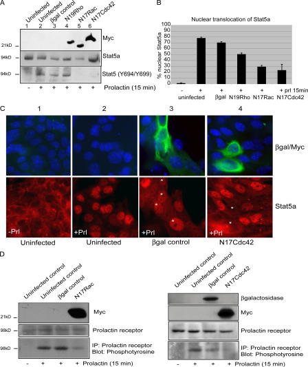
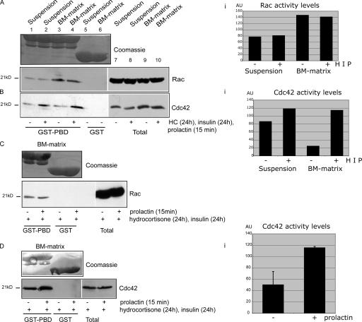
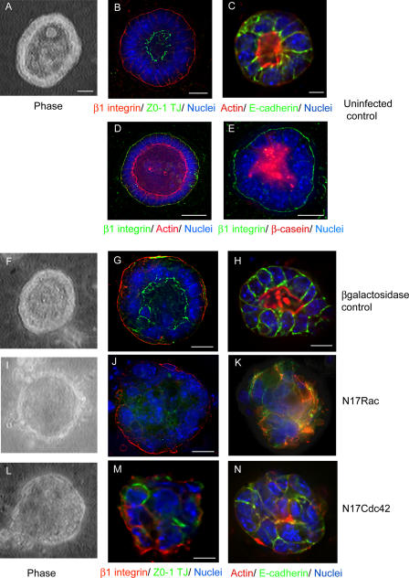
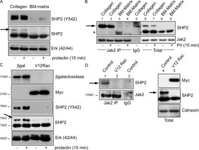
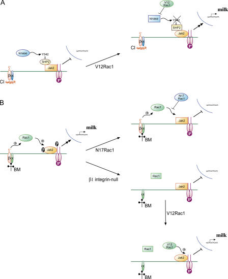
The expression of tissue-specific genes during mammary gland differentiation relies on the coincidence of two distinct signaling events: the continued engagement of beta1 integrins with the extracellular matrix (ECM) and a hormonal stimulus from prolactin (Prl). How the integrin and Prl receptor (PrlR) systems integrate to regulate milk protein gene synthesis is unknown. In this study, we identify Rac1 as a key link. Dominant-negative Rac1 prevents Prl-induced synthesis of the milk protein beta-casein in primary mammary epithelial cells cultured as three-dimensional acini on basement membrane. Conversely, activated Rac1 rescues the defective beta-casein synthesis that occurs under conditions not normally permissive for mammary differentiation, either in beta1 integrin-null cells or in wild-type cells cultured on collagen. Rac1 is required downstream of integrins for activation of the PrlR/Stat5 signaling cascade. Cdc42 is also necessary for milk protein synthesis but functions via a distinct mechanism to Rac1. This study identifies the integration of signals provided by ECM and hormones as a novel role for Rho family guanosine triphosphatases.


Figures

Figure 1.
Figure 1.
Rac and Cdc42 are essential for milk protein expression. (A) MECs were infected with Ad–β-galactosidase (lane 4), Ad-mycN19RhoA (lane 5), Ad-mycN17Rac1 (lane 6), Ad-mycN17Cdc42 (lane 7), or were not infected (lanes 1–3) and plated onto BM matrix for 24 h to assemble into 3D acini. Cultures were stimulated with Prl in differentiation medium for a further 24 h (lanes 2–7) or left untreated (lane 1), lysed, and immunoblotted with β-casein, β-galactosidase, or myc antibodies. Equal loading of protein in each lane was determined by levels of Erk. (B) As in A, but acini were stained for β-casein (red) and nuclei (blue). Micrographs are from sections taken midway through the spherical acinar structure. Bar, 7 μm.
Figure 2.
Figure 2.
Rac and Cdc42 regulate signal transduction through PrlR and Stat5. (A) MECs were uninfected (lanes 1 and 2) or infected with adenovirus as shown and plated onto BM matrix. 24 h later, acini were serum starved in differentiation medium for 24 h, stimulated with Prl for 15 min (lanes 2–6) or left untreated (lane 1), lysed, and immunoblotted with phospho-Stat5a/b (Y694/Y699) antibody or myc antibody. Total levels of Stat5 were determined by stripping and reprobing with an anti-Stat5a antibody. (B and C) Nuclear translocation of Stat5a was determined in virus-infected monolayer cells, and BM matrix was added to the medium (i.e., 2D culture with BM overlay). Cells were stimulated with Prl for 15 min or left untreated, fixed, and stained with an anti-Stat5a antibody and an anti-myc or anti–β-galactosidase antibody as described. (B) Percentage of cells displaying nuclear Stat5a (mean of six counts from two separate experiments). Error bars represent SD. (C) Representative micrographs. In noninfected cells, Stat5a translocates to the nucleus in response to Prl (compare panels 1 with 2); in Ad–β-galactosidase–infected cells (green, panel 3), Stat5a still translocates. However, the expression of N17Cdc42 inhibits Stat5a translocation (green, panel 4); similar images were obtained with N17Rac1 (not depicted). Asterisks represent infected cells. (D) MECs infected with adenovirus and plated onto BM matrix as in A. Tyrosine phosphorylation of PrlR was analyzed by immunoprecipitation with an anti-PrlR antibody followed by immunoblotting with 4G10. Total levels of PrlR was determined by blotting with anti-PrlR antibody, and viral infection was confirmed with anti– β-galactosidase or anti-myc antibodies.
Figure 3.
Figure 3.
Rac activity is regulated by BM matrix, whereas lactogenic hormones regulate Cdc42. (A and B) Rac (A) and Cdc42 (B) activity were analyzed using recombinant GST–Pak-binding domain (PBD) in pull-down assays of lysates from 3D acini plated on growth factor–reduced BM matrix (for 4 d) or cells placed in suspension (for 2 h). Free GST was used in controls to confirm specificity. MECs were cultured in differentiation medium with or without hydrocortisone and insulin continuously and Prl for 15 min. The Coomassie stain demonstrates that equivalent levels of GST-PBD and free GST were used in each sample. Lanes 1–4 were scanned, and the relative levels of Rac and Cdc42 activity were plotted in Ai and Bi. HIP; hydrocortisone, insulin, and Prl. (C and D) Rac (C) and Cdc42 (D) activity was assessed in 3D acini cultured in differentiation medium containing hydrocortisone and insulin and stimulated with Prl (15 min) or left untreated. Prl treatment increased Cdc42 activity but not Rac activity. The relative level and Cdc42 activity is plotted in Di. Error bars represent SD. AU, arbitrary units. White lines indicate that intervening lanes have been spliced out.
Figure 4.
Figure 4.
N17Rac and -Cdc42 disrupt the morphogenesis of 3D acini. (A–E) Cells plated on BM matrix form polarized 3D acini. (A) Phase-contrast micrograph of uninfected MECs showing cells at the periphery of an acinus and a central lumen. (B and C) 2-d-old polarized acini stained for ZO-1 (green) and β1 integrin (red; B) or actin (red) and E-cadherin (green; C). (D) 4-d-old acinus with expanded lumen stained for actin (red) and β1 integrin (green). (E) Prl-stimulated acinus showing β-casein (red) secreted into the lumen and β1 integrin (green) at the basal domain. Low-power views of some of these panels to show more acini are presented in Fig. S2 (available at http://www.jcb.org/cgi/content/full/jcb.200601059/DC1). (F–N) MECs were infected with Ad–β-galactosidase (F–H), Ad-mycN17Rac1 (I–K), or Ad-mycN17Cdc42 (L–N) viruses for 1 h in suspension and were plated onto BM matrix–coated coverslips for 48 h. 3D acini were stained for β1 integrin (red) and ZO-1 (green; G, J, and M) or E-cadherin (green) and actin (red; H, K, and N). Phase-contrast micrographs show the absence of lumen in N17Rac1 and N17Cdc42 acini (I and L, respectively) but not in controls (F). Nuclei are stained blue. Virus infection was confirmed by immunostaining with anti– β-galactosidase antibody or anti-myc antibody (not depicted). Micrographs are sections taken midway through acini. Bar (D),10 μM; (A–C, E, G, H, J, and M) 7 μM.
Figure 5.
Figure 5.
Rac and Cdc42 regulate differentiation through distinct mechanisms. Preassembled polarized 3D acini with lumens that formed after plating on BM matrix for 48 h were left untreated (A and F) or were infected directly with Ad-mycN17Cdc42 (B, D, G, and J) or Ad-mycN17Rac1 (C, E, H, I, K, and L). Acini were stained 24 h later for β1 integrin (red) and actin (green; A–C). Short arrows indicate cells collapsing into the lumen; long arrows indicate lumens. For milk studies, acini were stimulated with Prl for 24 h and stained for β-casein (red; F–I). N17Cdc42 did not inhibit β-casein synthesis, as indicated by immunofluorescence and blotting (G and M, respectively). N17Rac1 blocked β-casein synthesis (H and I). In I and L, only part of the acinus expressed N17Rac1 (dashed lines); only these cells failed to lactate. Viral infection was confirmed with myc antibody (D, E, and J–L). Bar, 7 μM.
Figure 6.
Figure 6.
V12Rac1 stimulates milk protein synthesis in cells cultured on nonpermissive collagen I substratum. (A) MECs were uninfected or infected with Ad-mycV12Rac1 and plated onto collagen I, where they formed 2D monolayers. Cells were stimulated with Prl for 24 h and were analyzed for β-casein expression by blotting. Infection was confirmed with myc or Rac antibody. These cultures were not overlaid with BM matrix, and it is well established that under these conditions, Prl cannot stimulate differentiation (lane 2). V12Rac1 rescued the defect (duplicate lanes 3 and 4). (B and C) Stat5A nuclear translocation was analyzed in uninfected cells or those infected with Ad-mycV12Rac1, plated onto collagen I–coated coverslips (2D culture with no BM matrix overlay), stimulated with Prl for 15 min, and stained with Stat5a and myc antibodies. (B) Percentage of cells displaying nuclear Stat5a; histogram is a mean of six counts from two experiments. Error bars represent SD. (C) Micrographs demonstrating the nuclear translocation of Stat5a (red) in cells expressing V12Rac1 (green) but not in uninfected controls. Note that monolayer-cultured MECs treated with BM overlay respond to Prl (Fig. 2 C), but in the experiment shown here, no exogenous BM is provided, and the cells cannot respond to Prl (ii). V12Rac1 rescues this defect (iv).
Figure 7.
Figure 7.
V12Rac1 restores the lactation defect observed in β1 integrin–null acini. (A) MECs from Itgβ1fx/fx mice were left untreated or were infected in monolayer with Cre-expressing adenovirus (Ad-CreM1) for 24 h and examined for β1 integrin by immunoblotting. (B) Cells treated as in A were reinfected with Ad–β-galactosidase (lanes 3 and 6) or Ad-mycV12Rac1 (lanes 4 and 7) in suspension for 1 h and were replated onto BM matrix for 24 h. 3D acini were untreated or stimulated with Prl for 24 h and analyzed for β-casein synthesis by immunoblotting. (C) Rac activity in 3D acini on BM matrix either uninfected or infected with Ad–β-galactosidase or Ad-CreM1. Rac activity was almost abolished in β1 integrin–null acini.
Figure 8.
Figure 8.
Phosphorylation of SHP2 is elevated in cells on collagen I and inhibited by V12Rac1. (A) MECs cultured in two dimensions on collagen I (no BM overlay) or as 3D acini on BM matrix were either untreated or stimulated with Prl for 15 min and immunoblotted for SHP2-pY542. The slower migrating band in the SHP2 blot (arrows in A–D) is a phosphorylated form of SHP2, which disappears after alkaline phosphatase treatment (not depicted) and comigrates with the position of SHP2-pY542. (B) Cell lysates prepared as in A were immunoprecipitated with Jak2 antibody (lanes 1–4) or rabbit IgG (lane 5) and immunoblotted with SHP2 antibody (top). Jak2 immunoblotting confirmed that equivalent levels were immunoprecipitated (bottom). Total levels of SHP2 and Jak2 are shown in lanes 6–9. Low levels of total SHP2 associate with Jak2 (asterisk), and SHP2-pY542 (arrow) only associates in the cells cultured on collagen I. The band in lane 5 is nonspecific and migrates more rapidly than SHP2. (C) MECs infected with Ad–β-galactosidase or Ad-V12Rac1 were plated on collagen I (no BM overlay) for 48 h and immunoblotted for SHP2-pY542 or total SHP2. V12Rac1 inhibits SHP2 phosphorylation. (D) Coimmunoprecipitation experiments show that phosphorylated SHP2 no longer complexes with Jak2 in the presence of V12Rac1. Jak2 immunoprecipitates (lanes 1 and 2) prepared from uninfected or V12Rac1-infected cells on collagen I were immunoblotted with SHP2 or Jak2 antibody. Expression of virus and total levels of SHP2 are shown in lanes 4 and 5.
Figure 9.
Figure 9.
Models for the involvement of Rac1 in MEC differentiation. (A) In MECs cultured on collagen I (CI), Prl (P) signaling is not active because the SHP2 associated with Jak2 is phosphorylated on Y542 by an unknown kinase. V12Rac1 suppresses this activity, leading to SHP2-Y542 dephosphorylation and Prl signaling. This builds on our previously proposed model for PTP involvement in MEC differentiation (Edwards et al., 1998). Active enzymes are oval, and inactive enzymes are rectangular. (B) In cells cultured on BM, Prl signaling requires Rac1. In either the presence of N17Rac1 or absence of β1 integrin, Rac1 activity is suppressed, leading to the inability of Prl to signal. V12 Rac1 can rescue Prl signaling in integrin-null MECs, but the mechanism may be different than that in cells on collagen I.

References

    1. Aggeler, J., J. Ward, L.M. Blackie, M.H. Barcellos-Hoff, C.H. Streuli, and M.J. Bissell. 1991. Cytodifferentiation of mouse mammary epithelial cells cultured on a reconstituted basement membrane reveals striking similarities to development in vivo. J. Cell Sci. 99:407–417. - PubMed
    1. Akhtar, N., and N.A. Hotchin. 2001. RAC1 regulates adherens junctions through endocytosis of E-cadherin. Mol. Biol. Cell. 12:847–862. - PMC - PubMed
    1. Barcellos-Hoff, M.H., J. Aggeler, T.G. Ram, and M.J. Bissell. 1989. Functional differentiation and alveolar morphogenesis of primary mammary cultures on reconstituted basement membrane. Development. 105:223–235. - PMC - PubMed
    1. Benitah, S.A., P.F. Valeron, H. Rui, and J.C. Lacal. 2003. STAT5a activation mediates the epithelial to mesenchymal transition induced by oncogenic RhoA. Mol. Biol. Cell. 14:40–53. - PMC - PubMed
    1. Benitah, S.A., M. Frye, M. Glogauer, and F.M. Watt. 2005. Stem cell depletion through epidermal deletion of Rac1. Science. 309:933–935. - PubMed

Publication types

MeSH terms