. 2006 Jul;4(7):e210.
doi: 10.1371/journal.pbio.0040210.

Translation repression in human cells by microRNA-induced gene silencing requires RCK/p54

Affiliations

Translation repression in human cells by microRNA-induced gene silencing requires RCK/p54

Chia-ying Chu et al. PLoS Biol. 2006 Jul.

Abstract

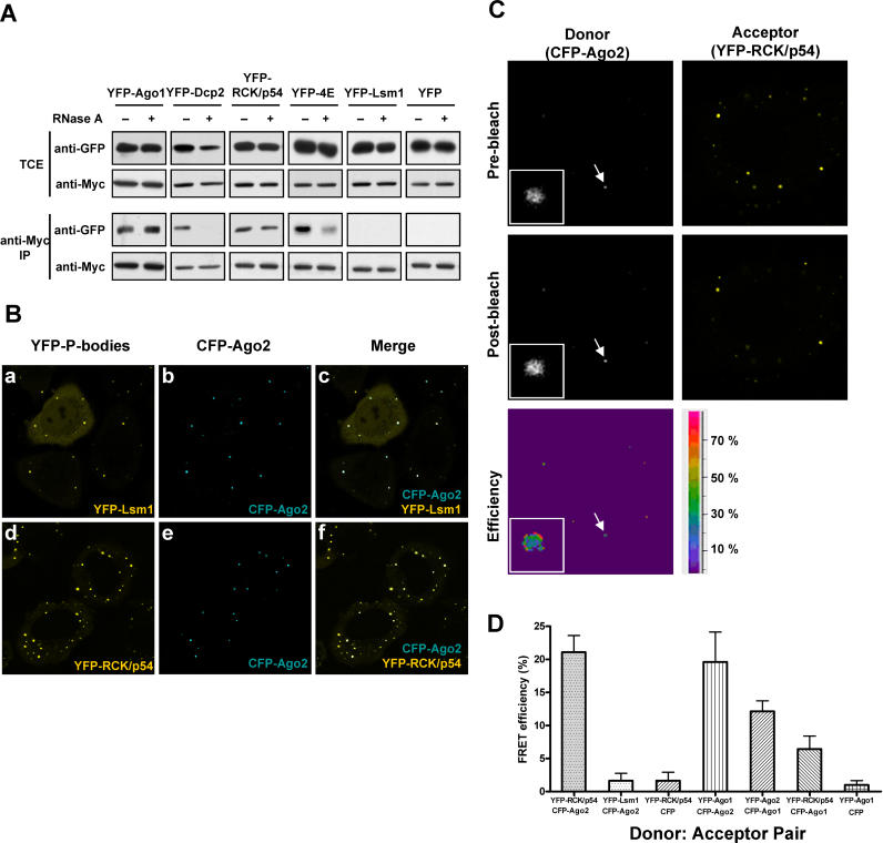
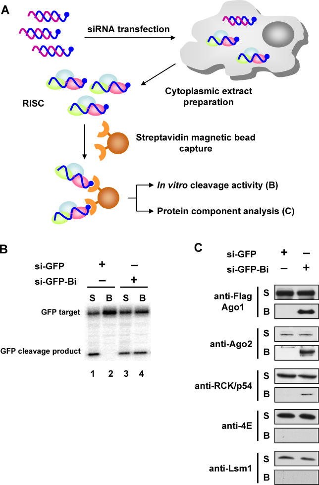
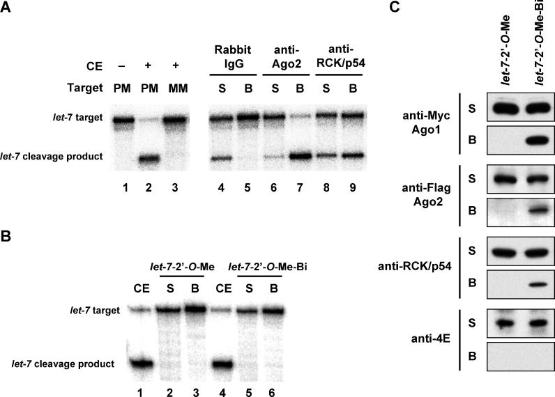
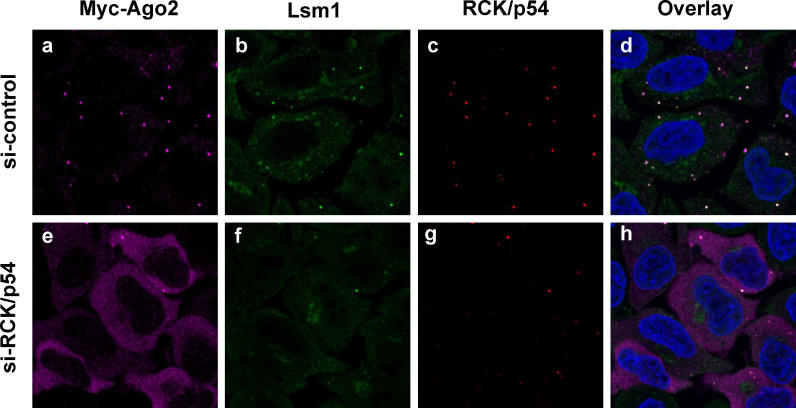
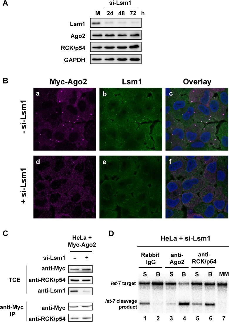
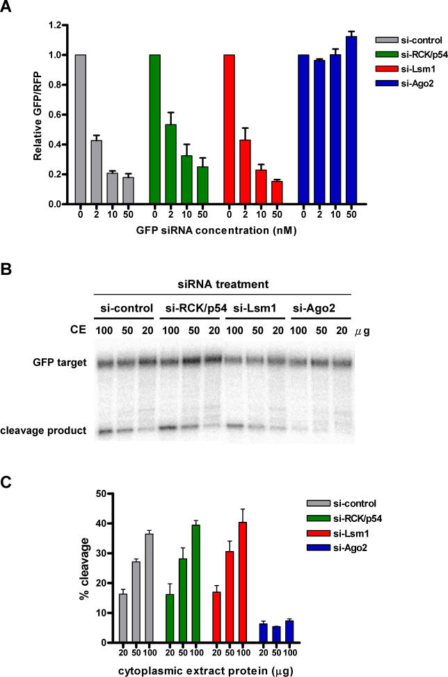
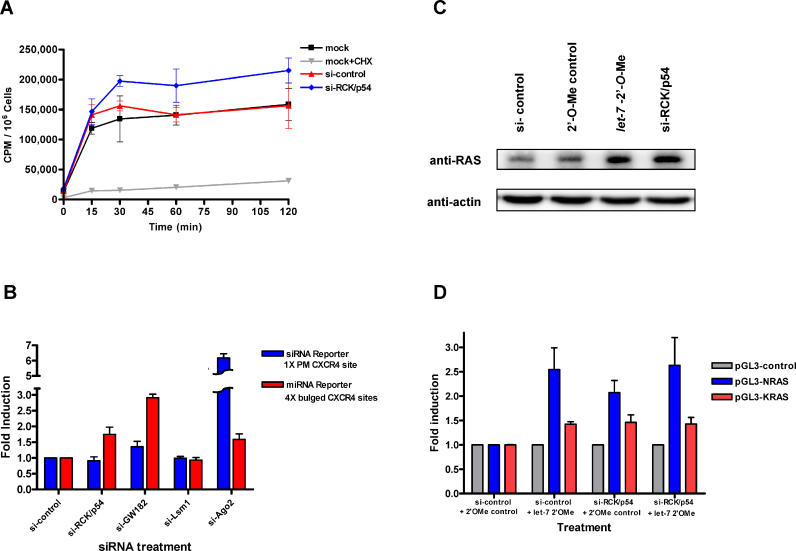
RNA interference is triggered by double-stranded RNA that is processed into small interfering RNAs (siRNAs) by Dicer enzyme. Endogenously, RNA interference triggers are created from small noncoding RNAs called microRNAs (miRNAs). RNA-induced silencing complexes (RISC) in human cells can be programmed by exogenously introduced siRNA or endogenously expressed miRNA. siRNA-programmed RISC (siRISC) silences expression by cleaving a perfectly complementary target mRNA, whereas miRNA-induced silencing complexes (miRISC) inhibits translation by binding imperfectly matched sequences in the 3' UTR of target mRNA. Both RISCs contain Argonaute2 (Ago2), which catalyzes target mRNA cleavage by siRISC and localizes to cytoplasmic mRNA processing bodies (P-bodies). Here, we show that RCK/p54, a DEAD box helicase, interacts with argonaute proteins, Ago1 and Ago2, in affinity-purified active siRISC or miRISC from human cells; directly interacts with Ago1 and Ago2 in vivo, facilitates formation of P-bodies, and is a general repressor of translation. Disrupting P-bodies by depleting Lsm1 did not affect RCK/p54 interactions with argonaute proteins and its function in miRNA-mediated translation repression. Depletion of RCK/p54 disrupted P-bodies and dispersed Ago2 throughout the cytoplasm but did not significantly affect siRNA-mediated RNA functions of RISC. Depleting RCK/p54 released general, miRNA-induced, and let-7-mediated translational repression. Therefore, we propose that translation repression is mediated by miRISC via RCK/p54 and its specificity is dictated by the miRNA sequence binding multiple copies of miRISC to complementary 3' UTR sites in the target mRNA. These studies also suggest that translation suppression by miRISC does not require P-body structures, and location of miRISC to P-bodies is the consequence of translation repression.


Figures

Figure 1
Figure 1. Human Argonaute Proteins Interact with RCK/p54, a Component of P-Bodies
(A) Immunoprecipitation and immunoblot analyses. TCEs from HeLa cells co-expressing Myc-Ago2 and YFP-Ago1, YFP-Dcp2, YFP-RCK/p54, YFP-eIF4E, YFP-Lsm1, or YFP were treated with +/− RNase A followed by Myc-Ago2 immunoprecipitation. TCE and anti-Myc IPs were analyzed by immunoblot using anti-GFP and anti-Myc antibodies. (B) In vivo localization of RCK/p54 and Ago2 to P-bodies. HeLa cells expressing YFP-Lsm1 and CFP-Ago2 (a, b, and c), YFP-RCK/p54 and CFP-Ago2 (d, e, and f) were visualized by confocal microscopy at 24 h post-transfection. (C) Visualization of interactions between RCK/p54 and Ago2 in P-bodies by FRET. HeLa cells expressing YFP-RCK/p54 and CFP-Ago2 were fixed at 24 h post-transfection. FRET was measured by an acceptor photobleaching method. Fluorescence images of donor (CFP-Ago2) and acceptor (YFP-RCK/p54) molecules were taken before and after photobleaching YFP. FRET efficiencies were calculated as described [ 48, 49, 68], and data were analyzed by Leica confocal software. Arrows point to P-bodies, which are enlarged in insets. (D) FRET efficiencies between different P-body protein donor: acceptor pairs. HeLa cells co-expressing YFP-RCK/p54 and CFP-Ago2, YFP-Lsm1 and CFP-Ago2, YFP-RCK/p54 and CFP, YFP-Ago1 and CFP-Ago2, YFP-Ago2 and CFP-Ago1, YFP-RCK/p54 and CFP-Ago-1, as well as YFP-Ago1 and CFP, were fixed and FRET efficiencies were measured.
Figure 2
Figure 2. Isolation of Active Human RISC Containing RCK/p54-Ago1/Ago2
(A) Experimental outline to purify active human RISC. The guide strands of siRNA complexes targeting GFP (si-GFP) were conjugated with 3′ biotin (si-GFP-Bi; blue strands) and transfected into HeLa cells. RISCs were captured by incubating cell extracts with streptavidin-magnetic beads. (B) Target mRNA is cleaved by biotin-captured RISC. Bead (B) and supernatant (S) phases of captured RISC were incubated with 124-nt 32P-cap-labeled GFP target mRNA. The reactions were stopped after 120 min, and products were resolved on 6% denaturing polyacrylamide gels. (C) Biotin-captured RISC contains proteins associated with mRNA processing. Active human RISC from HeLa cells expressing Flag-Ago1 was captured by biotin-siRNA and its protein composition was analyzed by immunoblot using anti-Flag, anti-Ago2, anti-RCK/p54, anti-Lsm1, and anti-eIF4E antibodies.
Figure 3
Figure 3. RCK/p54 Is a Component of Human miRISC
(A) Affinity-purified miRISCs associated with PCK/p54 retain cleavage activity. To purify miRISC associated with RCK/p54, magnetic protein A beads coupled with rabbit IgG, rabbit anti-Ago2, or rabbit anti-RCK/p54 antibodies were incubated with HeLa cytoplasmic extracts. After immunoprecipitation, RISC activities were analyzed by incubating the supernatant (S) or bead (B) phases with 182-nt 32P-cap-labeled let-7 substrate mRNAs having a perfectly complementary or mismatched sequence to the let-7 miRNA. Cleavage products were resolved on 6% denaturing polyacrylamide gels. CE, cytoplasmic extract; PM, perfect match; MM, mismatch. (B) Affinity-purified miRISCs retain cleavage activity. let-7 miRISC cleavage of a perfectly matched RNA target was inhibited by 2′- O-Me oligonucleotides complementary to let-7 miRNA ( let-7–2′- O-Me or let-7–2′- O-Me-biotin). A 182-nt 32P-cap-labeled let-7 substrate mRNA was incubated with the supernatant (S) or bead (B) phases of captured miRISC. The reactions were stopped after 120 min, and products were resolved on 6% denaturing polyacrylamide gels. (C) miRISCs contain proteins associated with mRNA processing. Cytoplasmic extracts of HeLa cells expressing Flag-Ago2 and Myc-Ago1 were incubated with 2′- O-Me oligonucleotides complementary to let-7 miRNA ( let-7–2′- O-Me or let-7–2′- O-Me-biotin), affinity-purified by streptavidin-magnetic beads to capture let-7 miRISC. Supernatant (S) and beads (B) after biotin capture were analyzed by immunoblot using anti-Myc, anti-Flag, anti-RCK/p54, and anti-eIF4E antibodies.
Figure 4
Figure 4. Depletion of RCK/p54 Disrupts P-bodies and Disperses the Localization of Human Ago2
HeLa cells were co-transfected with Myc-Ago2 and siRNA against human RCK/p54 (lower panels) or CDK9 mismatch (control; upper panels). At 24 h post-transfection, cells were analyzed by immunofluorescence using antibodies against Myc-Ago2 (A and E) and against the P-body proteins Lsm1 (B and F) and RCK/p54 (C and G). Cells were stained with Hoechst 33258 to visualize nuclei and images were digitally merged (D and H).
Figure 5
Figure 5. Depletion of Lsm1 Disrupts P-Bodies but Does Not Affect the Interaction between RCK/p54 and Ago2
(A) Specific knockdown of Lsm1 in HeLa cells by siRNA. HeLa cells were transfected with siRNA against Lsm1 and harvested at 24, 48, and 72 h post-transfection, and TCEs were analyzed by immunoblot with antibodies against Lsm1 or GAPDH. (B) Depletion of Lsm1 disrupts P-bodies. HeLa cells were transfected with siRNA against Lsm1. At 48 h post-transfection, cells were analyzed by immunofluorescence using antibodies against Lsm1 and Myc tag for Ago2. Cells were stained with Hoechst33258 to visualize nuclei, and images were digitally merged. (C) RCK/p54 interacts with Myc-Ago2 in Lsm1-depleted cells. HeLa cells were transfected for 48 h with Myc-Ago2 and control siRNA or siRNA against Lsm1, TCEs were prepared, and Myc-Ago2 was immunoprecipitated from an aliquot of TCE. TCE and anti-Myc IPs were analyzed by immunoblot using anti-Myc, anti-RCK/p54, and anti-Lsm1 antibodies. (D) Affinity-purified RCK/p54 and Ago2 from Lsm1-depleted cell extracts retain miRISC activity. HeLa cells were transfected for 48 h with siRNA against Lsm1, and cytoplasmic extracts were prepared. These extracts were incubated with magnetic protein A beads coupled with rabbit IgG, rabbit anti-Ago2, or rabbit anti-RCK/p54 antibodies to purify miRISC associated with RCK/p54. After immunoprecipitation, RISC activities were analyzed by incubating the supernatant (S) or bead (B) phases with 182-nt 32P-cap-labeled let-7 substrate mRNAs having a perfectly matched or a mismatched sequence to the let-7 miRNA. Cleavage products were resolved on 6% denaturing polyacrylamide gels. MM, mismatch.
Figure 6
Figure 6. Depletion of RCK/p54 Has No Significant Effect on RNAi Activity In Vivo and In Vitro
(A) In vivo effect of depleting RCK/p54, Lsm1, and Ago2 on siRNA dose-dependent RNAi activity. HeLa cells were transfected with siRNAs against CDK9 mismatch (control), RCK/p54, Lsm1, or Ago2. 24 h later cells were transfected again with EGFP and RFP reporter plasmids and varying amounts (2, 10, and 50 nM) of siRNA against EGFP. 24 h after the second transfection, RNAi efficiencies were analyzed (see Materials and Methods). To quantify the effect of depleting RCK/p54 and Ago2 on RNAi, the ratio of GFP/RFP signals was normalized to that observed in the absence of GFP siRNA (0 nM). Normalized ratios < 1.0 indicate specific RNAi at a given siRNA concentration. (B) Depletion of RCK/p54 has no effect on in vitro siRISC cleavage activity. HeLa cells were transfected with siRNAs targeting RCK/p54, Lsm1, Ago2, or CDK9 mismatch (control). 24 h after the first transfection, cells were again transfected with 50 nM siRNA targeting EGFP. 24 h later cytoplasmic extracts were made (see Materials and Methods), and varying amounts (20, 50, 100 μg) of total cytoplasmic extract protein were incubated with a 124-nt, 32P-cap-labeled GFP target mRNA. The reactions were stopped after 60 min, and products were resolved on 6% denaturing polyacrylamide gels. (C) Quantification of siRISC cleavage activity in vitro after depletion of RCK/p54, Lsm1, or Ago2. Cleavage activity of siRISC targeting EGFP mRNA was quantified as a function of protein content in extracts of HeLa cells depleted of RCK/p54, Lsm1, or Ago2.
Figure 7
Figure 7. RCK/p54 Represses General and miRNA-Mediated Translation
(A) Release of general translational repression by silencing RCK/p54. Incorporation of [ 35S]methionine into HeLa cells was used to measure general translational activity. Cells were transfected with 50 nM siRNAs targeting mismatched CDK9 (control) or RCK/p54. Mock control cells were treated with the transfection reagent only. At 24 h post-transfection, cells were incubated for 1 h in medium lacking Met and Cys, and metabolically labeled with [ 35S]methionine (see Materials and Methods). As a control for passive uptake of [ 35S], mock cells were treated with 40 μg/ml of the translation inhibitor, cycloheximide. Cell incorporation of [ 35S] is shown as cpm/10 6 cells versus time after adding [ 35S]. (B) Depletion of RCK/p54 releases translational repression in a luciferase reporter system. HeLa cells were transfected with siRNAs against CDK9 mm (control), RCK/p54, GW182, Lsm1, or Ago2. 24 h later, cells were co-transfected with siRNA (1 × perfectly matched [PM] site) or miRNA (4 × bulged sites) luciferase reporters in the presence of CXCR4 siRNA. RL activities were measured 24 h after the second transfection and normalized to FL activity as a control. Repression of reporter gene expression by siRNA or miRNA were measured by normalizing the RL/FL signals in the presence of CXCR4 siRNA to the RL/FL activities observed in the absence of siRNA. Release of gene repression by siRNA or miRNA is presented as the fold induction of RL/FL activities compared to the RL/FL signals observed in control (si-CDK9mm) experiments. FL, firefly luciferase. (C) Depletion of RCK/p54 releases repression of RAS protein translation. HeLa cells were transfected with 100 nM of 2′- O-Me oligonucleotide ( let-7 2′- O-Me inhibitor or 2′- O-Me control), and siRNA against RCK/p54 or CDK9 mm control. 24 h after transfection, the cells were counted and harvested (see Materials and Methods). The protein contents of TCEs were resolved by SDS-PAGE and analyzed by immunoblot using anti-RAS and anti-actin antibodies. (D) Depletion of RCK/p54 releases repression of luciferase protein translation by 3′ UTRs of NRAS and KRAS. HeLa cells transfected with an Rr-luc-expressing vector, pRL-TK, and a Pp- luc-expressing vector, pGL3-control, pGL3-NRAS, or pGL3-KRAS, were co-transfected with 100 nM of 2′- O-Me oligonucleotide ( let-7 2′- O-Me inhibitor or 2′- O-Me control) and siRNA against RCK/p54 or CDK9 mm control. TCEs were prepared 48 h post-transfection and dual luciferase assays were performed. Relative GL ( Pp-luc)/RL ( Rr-luc) signals were normalized to those of pGL3-control-transfected cells and are shown as the fold induction compared to mock treatment.
Figure 8
Figure 8. A Model for Human RISC Function Involving miRNA and siRNA
RISC contains Ago2 (red), Ago1 (green), RCK/p54 (blue, labeled p54), and other known (e.g., Dicer and TRBP) and unidentified proteins (pink) and is distributed throughout the cytoplasm. siRISC binds to its target mRNA by perfectly matching base pairs, cleaves the target mRNA for degradation, recycles the complex, and does not require P-body structures for its function. Multiple numbers (n) of miRISC bind to target mRNA by forming a bulge sequence in the middle that is not suitable for RNA cleavage, accumulate in P-bodies, and repress translation by exploiting global translational suppressors such as RCK/p54. The translationally repressed mRNA is either stored in P-bodies or enters the mRNA decay pathway for destruction. Depending upon cellular conditions and stimuli, stored mRNA can either re-enter the translation or mRNA decay pathways.

Comment in

References

    1. Wienholds E, Plasterk RH. MicroRNA function in animal development. FEBS Lett. 2005;579:5911–5922. - PubMed
    1. Ambros V. The functions of animal microRNAs. Nature. 2004;431:350–355. - PubMed
    1. Bartel DP. MicroRNAs: Genomics, biogenesis, mechanism, and function. Cell. 2004;116:281–297. - PubMed
    1. Filipowicz W. RNAi: The nuts and bolts of the RISC machine. Cell. 2005;122:17–20. - PubMed
    1. Fire A, Xu S, Montgomery MK, Kostas SA, Driver SE, et al. Potent and specific genetic interference by double-stranded RNA in Caenorhabditis elegans . Nature. 1998;391:806–811. - PubMed

Publication types

MeSH terms