. 2006 Jun 13;103(24):9023-8.
doi: 10.1073/pnas.0602681103. Epub 2006 Jun 6.

GPR56, an atypical G protein-coupled receptor, binds tissue transglutaminase, TG2, and inhibits melanoma tumor growth and metastasis

Affiliations

GPR56, an atypical G protein-coupled receptor, binds tissue transglutaminase, TG2, and inhibits melanoma tumor growth and metastasis

Lei Xu et al. Proc Natl Acad Sci U S A. .

Abstract

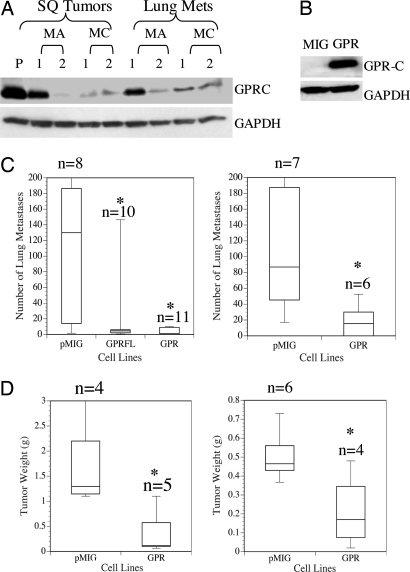
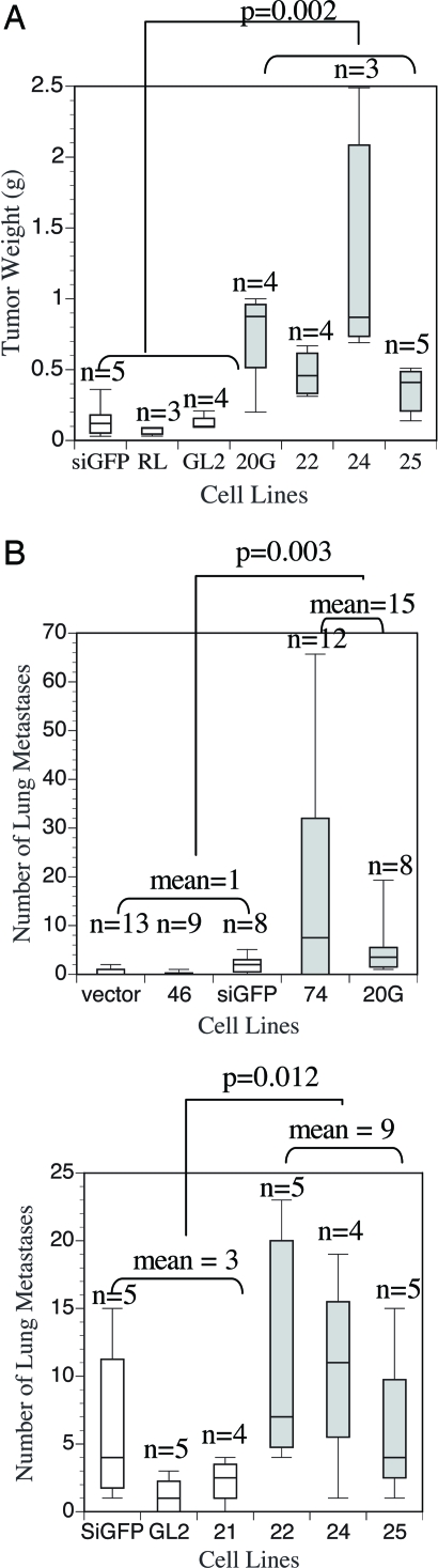
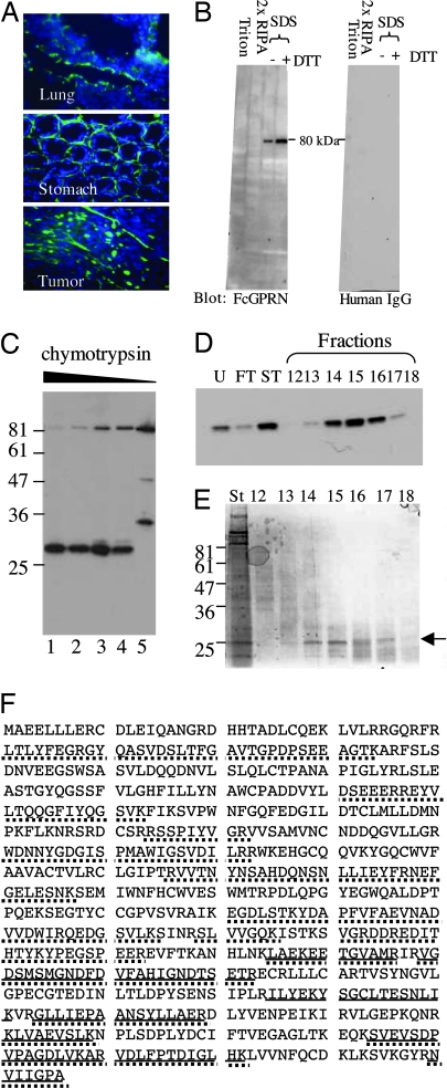
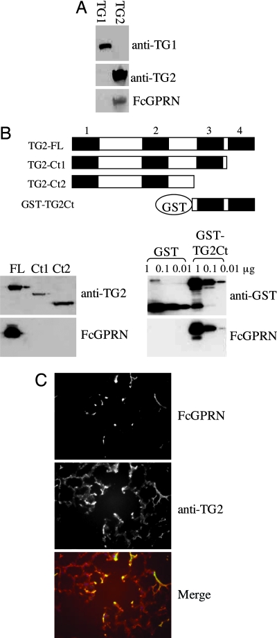
The survival and growth of tumor cells in a foreign environment is considered a rate-limiting step during metastasis. To identify genes that may be essential for this process, we isolated highly metastatic variants from a poorly metastatic human melanoma cell line and performed expression analyses of metastases and primary tumors from these cells. GPR56 is among the genes markedly down-regulated in the metastatic variants. We show that overexpression of GPR56 suppresses tumor growth and metastasis, whereas reduced expression of GPR56 enhances tumor progression. Levels of GPR56 do not correlate with growth rate in vitro, suggesting that GPR56 may mediate growth suppression by interaction with a component in the tumor microenvironment in vivo. We show that GPR56 binds specifically to tissue transglutaminase, TG2, a widespread component of tissue and tumor stroma previously implicated as an inhibitor of tumor progression. We discuss the mechanisms whereby GPR56-TG2 interactions may suppress tumor growth and metastasis.


Conflict of interest statement

Conflict of interest statement: No conflicts declared.

Figures

Fig. 1.
Fig. 1.
GPR56 is down-regulated in tumors from metastatic variants, and its reexpression suppresses tumor growth and metastasis in MC-1 cells. (A) Expression of GPR56 was examined by immunoblotting lysates of tumors from all of the cell lines by using anti-GPRC antibody. GAPDH was used as a loading control. GPR56 protein levels were reduced in both s.c. tumors and lung metastases derived from highly metastatic melanoma cells as compared with s.c. tumors of the poorly metastatic parental line (P). (B) GPR56 was expressed in MC-1 cells as determined by anti-GPR56 antibody on a Western blot. (C) Experimental metastasis assays of MC-1 cells with empty vector (pMIG) or expressing GPR56 (GPR) or FLAG-tagged GPR56 (GPRFL). Cells (5 × 10⁵) were injected intravenously, and lung metastases were counted 2 months later. MC-1(pMIG-GPR) or MC-1(pMIG-GPRFL) cells produced significantly fewer metastases (two independent experiments). ∗, P < 0.05. (D) MC-1 cells expressing high levels of GPR56 also show significantly reduced primary tumor growth. Weights of s.c. tumors from MC-1(pMIG-GPR) or MC-1(pMIG) cells are shown. ∗, P < 0.05.
Fig. 2.
Fig. 2.
Reduction of GPR56 by RNAi promotes tumor growth and metastasis. (A) Reduction of GPR56 resulted in increased s.c. tumor growth in vivo. Weights of s.c. tumors from A375-RNAi cells are shown. Box plots of A375-RNAi cells with significant reduction of GPR56 protein (see Fig. 8) are shaded. Statistical significance is shown for all of these A375-RNAi lines versus controls. (B) Reduction of GPR56 resulted in an enhancement of metastasis in vivo as shown by experimental metastasis assays. A375eco-RNAi cells (5 × 10⁵) or controls were injected intravenously into immunodeficient nude mice (Upper) and nonobese diabetic severe combined immunodeficient (NOD-SCID) mice (Lower). Lung metastases were counted 2 months later. Box plots of A375eco-RNAi cells with significant reduction of GPR56 protein (see Fig. 8) are shaded. Statistical significance is shown for all of these A375-RNAi lines versus controls.
Fig. 3.
Fig. 3.
Identification of TG2 as a candidate GPR56-binding protein. (A) The N terminus of GPR56 that was fused to human Fc fragment (FcGPRN) recognizes extracellular matrix on tissue and tumor sections by immunohistochemistry. Blue, DAPI; green, FcGPRN. (B) FcGPRN recognized a band of ≈80 kDa in the radioimmunoprecipitation assay (RIPA)-insoluble fraction of lung lysate. The extraction scheme is shown in Fig. 9. Human IgG was used as a negative control. (C) FcGPRN recognizes an ≈28-kDa fragment after chymotryptic digestion of mouse lungs. Lanes 1–4 are 1:100, 1:250, 1:500, and 1:1,000 dilutions of 2.5% chymotrypsin. Lane 5 is a negative control with no chymotrypsin treatment. (D) The fractions after purification (indicated in Fig. 10) were probed by FcGPRN on a Western blot. (E) Some of the samples shown in D were stained with SimplyBlue stain and the ≈28-kDa bands were excised for mass spectrometric analysis (arrow). (F) Identification of mouse TG2 as a candidate protein from mass spectrometric analysis. The sequence of TG2 protein is shown. Bold lines indicate the sequences found in the ≈28-kDa fragment, and the dotted lines indicate the sequences found in the uncleaved ≈80-kDa protein. The ≈28-kDa fragment is located at the C terminus of the protein.
Fig. 4.
Fig. 4.
TG2 is a binding partner of GPR56. (A) FcGPRN recognizes murine TG2, but not TG1, from transfected 293T cells. The 293T cells were transiently transfected with murine TG1 or TG2 in pCMV-SPORT6 vector for 48 h, and total cell lysates were run on polyacrylamide gels and probed with anti-TG1 antibody, anti-TG2 antibody, or FcGPRN. (B) The C-terminal TG2 domains are both necessary and sufficient for the binding of GPR56. The TG2 protein and its fragments tested for FcGPRN binding are schematically shown. The four conserved domains are presented as four black boxes and numbered as 1–4. Domain 1, N-terminal β-sandwich domain; domain 2, catalytic core; domains 3 and 4, the two C-terminal β-barrel domains. The sequences between domains are shown as white boxes. TG2-FL, full-length TG2; TG2-Ct1, TG2 protein lacking the last β-barrel domain; TG2-Ct2, TG2 protein lacking the last two β-barrel domains; and GST-TG2Ct, the last two β-barrel domains were expressed in E. coli as a GST fusion protein. FcGPRN binds to the C terminus of TG2 as well as the full-length TG2 but not to TG1 or truncated TG2 lacking C-terminal domain(s). The C terminus of TG2 is also sufficient to mediate the binding between TG2 and FcGPRN. (C) FcGPRN and anti-TG2 antibody show overlapping staining patterns on lung sections. In the merged picture, green is from FcGPRN staining, and red is from anti-TG2 staining.

References

    1. Fidler I. J. Nat. Rev. Cancer. 2003;3:453–458. - PubMed
    1. MacDonald I. C., Groom A. C., Chambers A. F. BioEssays. 2002;24:885–893. - PubMed
    1. Clark E. A., Golub T. R., Lander E. S., Hynes R. O. Nature. 2000;406:532–535. - PubMed
    1. Kang Y., Siegel P. M., Shu W., Drobnjak M., Kakonen S. M., Cordon-Cardo C., Guise T. A., Massague J. Cancer Cell. 2003;3:537–549. - PubMed
    1. Li C., Wong W. H. Proc. Natl. Acad. Sci. USA. 2001;98:31–36. - PMC - PubMed

Publication types

Substances