. 2006 Sep 15;108(6):2106-13.
doi: 10.1182/blood-2006-03-007427. Epub 2006 Jun 6.

Host MHC class II+ antigen-presenting cells and CD4 cells are required for CD8-mediated graft-versus-leukemia responses following delayed donor leukocyte infusions

Affiliations

Host MHC class II+ antigen-presenting cells and CD4 cells are required for CD8-mediated graft-versus-leukemia responses following delayed donor leukocyte infusions

Ronjon Chakraverty et al. Blood. .

Abstract

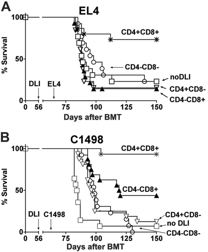
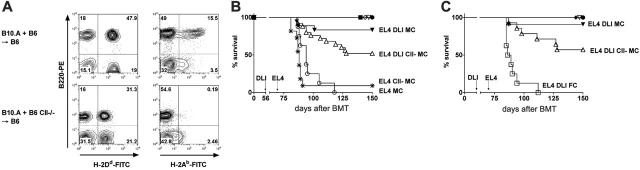
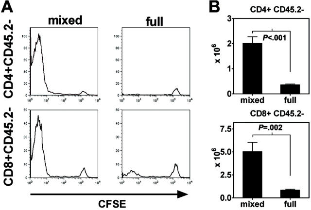
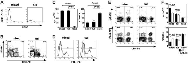
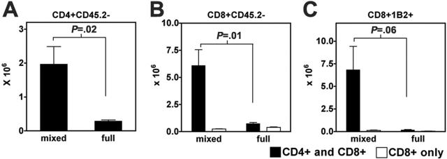
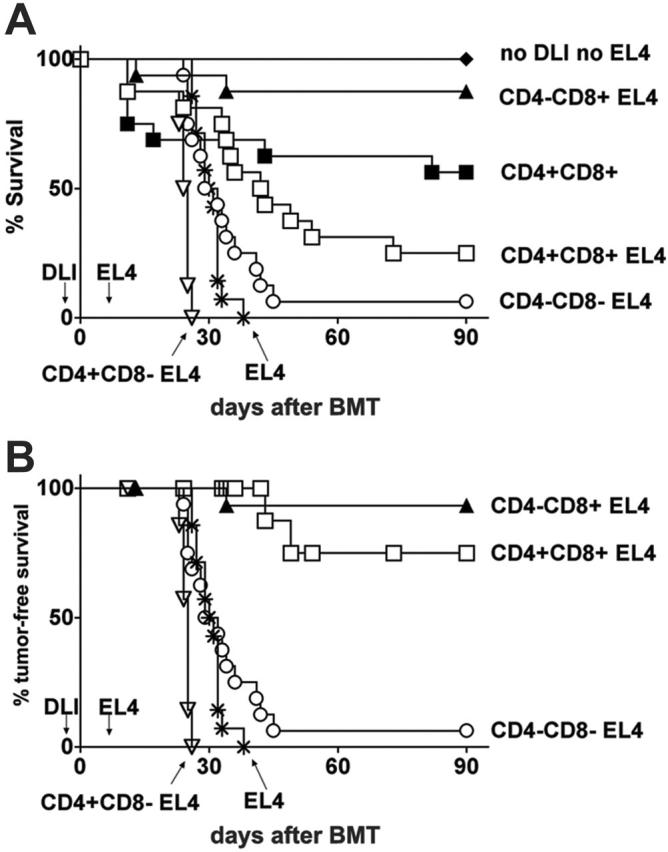
Following bone marrow transplantation, delayed donor leukocyte infusions (DLIs) can induce graft-versus-leukemia (GVL) effects without graft-versus-host disease (GVHD). These antitumor responses are maximized by the presence of host hematopoietic antigen-presenting cells (APCs) at the time of DLI. Using a tumor-protection model, we demonstrate here that GVL activity following administration of DLIs to established mixed chimeras is dependent primarily on reactivity to allogeneic MHC antigens rather than minor histocompatibility or tumor-associated antigens. CD8(+) T-cell-dependent GVL responses against an MHC class II-negative tumor following delayed DLI require CD4(+) T-cell help and are reduced significantly when host APCs lack MHC class II expression. CD4(+) T cells primed by host APCs were required for maximal expansion of graft-versus-host reactive CD8(+) T cells but not their synthesis of IFN-gamma. In contrast, the GVL requirement for CD4(+) T-cell help was bypassed almost completely when DLI was administered to freshly irradiated recipients, indicating that the host environment is a major factor influencing the cellular mechanisms of GVL.


Figures

Figure 1.
Figure 1.
GVL effects of delayed DLI. (A) B10.A → B6 FCs received 3 × 10⁷ B10.A DLI alone (▴, n = 7) on day 56 after BMT, C1498 myeloid leukemia cells (5 × 10⁴) alone (*, n = 8) on day 63, or both (□, n = 8). (B) In the same experiment, B10.A + B6 → B6 MCs received DLI alone (•, n = 6) on day 56, C1498 cells alone (○, n = 7) on day 63, or both (▾, n = 7). MCs receiving DLI and C1498 (▾, n = 7) showed markedly improved survival compared with FCs receiving DLI and C1498 (panel A: □, n = 8; P < .01). (C) C3.SW → B6 FCs received 1 × 10³ EL4 cells (*, n = 7) on day 63 or 3 × 10⁷ C3.SW SCs as DLI on day 56 after BMT plus 1 × 10³ EL4 on day 63 (□, n = 11). (D) C3.SW + B6 → B6 MCs also received EL4 cells alone (○, n = 7) on day 63, or both DLI and EL4 cells (▾, n = 10). MCs receiving DLI and EL4 showed similar survival compared with full chimeras receiving DLI and EL4.
Figure 2.
Figure 2.
Both CD4+ and CD8+ T cells are required for maximal GVL effects of delayed DLI in MCs. Data were pooled from 2 independent experiments. (A) B10.A + B6 → B6 MCs received 3 × 10⁷ nondepleted CD4+CD8+ DLIs (*, n = 15; inoculum contained a total of 5.2 × 10⁶ CD4+ plus 2.5 × 10⁶ CD8+ T cells), CD4CD8+ DLI (▴, n = 14; inoculum contained 2.5 × 10⁶ CD8+ T cells), CD4+CD8 DLI (▿, n = 13; inoculum contained 5.2 × 10⁶ CD4+ T cells), CD4CD8 DLI (○, n = 13), or no DLI (□, n = 13) on day 56 after BMT. All mice received 1 × 10³ EL4 T leukemia cells on day 63. (B) MCs received 3 × 10⁷ B10.A nondepleted CD4+CD8+ DLIs (*, n = 16; inoculum contained a total of 5.7 × 10⁶ CD4+ plus 2.3 × 10⁶ CD8+ T cells), CD4CD8+ DLI (▴, n = 16; inoculum contained 2.3 × 10⁶ CD8+ T cells), CD4+CD8 DLI (▿, n = 16; inoculum contained 5.8 × 10⁶ CD4+ T cells), CD4CD8 DLI (○, n = 13), or no DLI (□, n = 14) on day 56 after BMT followed by 5 × 10⁴ C1498 myeloid leukemia cells on day 63.
Figure 3.
Figure 3.
MHC class II expression by bone marrow–derived APCs is required for maximal GVL effects in MCs. (A) Peripheral blood chimerism analysis was performed in various cell lineages by flow cytometry 6 weeks after BMT. Representative contour plots show B10.A donor MHC class I (34-2-12, H2-Dd) and B6 recipient MHC class II (H2-Ab) expression in B cells of MCs generated by reconstitution with B10.A and either B6 MHC class II+/+ or B6 MHC class II–/– bone marrow. (B) MCs received either 3 × 10⁷ B10.A DLIs on day 56 after BMT, EL4 on day 63, or both. B6 MHC class II+/+ plus B10.A MCs: no DLI (▪, n = 4), DLI (•, n = 7), DLI + EL4 (▾, n = 18), EL4 (○, n = 8). B6 MHC class II–/– plus B10.A MCs (CII MCs): no DLI (⋄, n = 3), DLI (▿, n = 5), DLI + EL4 (▵, n = 25), EL4 (*, n = 11). Data were pooled from 2 similar, independent experiments. (C) Comparisons of GVL activities of DLI in MCs versus CII MCs versus FCs. MCs: DLI (• n = 7), DLI + EL4 (▾, n = 11). CII MCs: DLI (▿, n = 2), DLI + EL4 (▵, n = 14). FCs: DLI (♦, n = 6), DLI + EL4 (□, n = 8).
Figure 4.
Figure 4.
Donor CD4+ and CD8+ T-cell expansion requires the presence of host-derived hematopoietic APCs. B6 CD45.1 SCs (1 × 10⁷) and 2C CD8+ T cells (1 × 10⁶) were CFSE-labeled and then transferred to B6 CD45.2 + BALB/c → BALB/c MCs or B6 CD45.2 → BALB/c FCs 10 weeks following BMT. (A) Histograms show log CFSE staining of gated CD4+CD45.2 and CD8+CD45.2 T-cell populations within the spleen on day +6 following DLI. Results are representative of 3 independent experiments. (B) Absolute numbers of DLI-derived CD4+ and CD8+ T cells in recipient spleens of MCs and FCs on day +12 following DLI (n = 6 each group). Data are shown as mean ± SEM.
Figure 5.
Figure 5.
Host hematopoietic APCs prime host-specific donor T-cell expansion. B6 CD45.1 SCs (1 × 10⁷) and 2C CD8+ T cells (1 × 10⁶) were CFSE-labeled and then transferred to B6 CD45.2 + BALB/c → BALB/c MCs or B6 CD45.2 → BALB/c FCs 10 weeks following BMT. (A) Histograms show log CFSE staining of gated CD8+1B2+ T-cell populations within the spleen on day +6 following transfer. 1B2 is a clonotypic marker that identifies cells bearing the 2C TCR. Data are representative of 3 independent experiments. (B) Representative flow cytometric contour plots of recipient spleens of MCs and FCs on day +12 following DLI (x-axis, CD8-PE; y-axis, 1B2-APC). Data are representative of 3 independent experiments. (C) Percentage of gated CD8+1B2+ T cells that were CD44high (left) or CD62L+ (right) in recipient spleens of MCs and FCs on day +12 following DLI (naive controls shown for comparison, n = 3 each group). Data shown represent 1 of 2 independent experiments. (D) Representative flow cytometric plots of intracellular staining of IFN-γ in gated CD8+1B2+ T cells in recipient spleens of MCs and FCs on day +12 following DLI after brief ex vivo stimulation. Percentages of gated CD8+1B2+ cells that were IFN-γ+ were 40% ± 9% in MCs versus 37% ± 8% in FCs (P = NS; mean ± SEM; n = 4 each group). (E) Representative flow cytometric contour plots of recipient spleens of MCs and FCs on day +12 following DLI on gated CD45.2 cells (x-axis, CD4-PE; y-axis, Vβ3-APC or Vβ8.1/2-APC). (F) Percentage of gated CD4+Vβ3+ or CD4+Vβ8.1/2+ T cells that were CD44high (top) or CD62L+ (bottom) in recipient spleens of MCs and FCs on day +12 following DLI (naive phenotype shown for comparison, n = 3 each group). (C,F) Data are shown as mean ± SEM.
Figure 6.
Figure 6.
Graft-versus-host reactive donor CD8+ T-cell expansion following delayed DLI is dependent on CD4+ T-cell help. B6 CD45.1 CD8+ T cells (1 × 10⁶) and 2C CD8+ T cells (1 × 10⁶) with or without 2 × 10⁶ B6 CD45.1 CD4+ T cells were transferred to B6 CD45.2 + BALB/c → BALB/c MCs or B6 CD45.2 → BALB/c FCs 10 weeks following BMT. (A-C) Histograms show absolute numbers of CD4+CD45.2, CD8+CD45.2, and CD8+1B2+ populations within the spleen on day 12 following transfer (n = 3 each group). Open bars indicate CD8+ T cells transferred without CD4+ T cells; closed bars, CD4+ and CD8+ T cells cotransferred. Note that in Figure 6A, only closed bars are shown (CD4+ and CD8+ T cells cotransferred). Data are shown as mean ± SEM.
Figure 7.
Figure 7.
CD4+ T cells do not contribute to GVL effects of DLI in freshly irradiated BMT recipients. Lethally irradiated B6 mice were reconstituted with a mixture of TCD B10.A and B6 bone marrow together with nondepleted DLI (□, n = 8; inoculum contained a total of 5.3 × 10⁵ CD4+ plus 2.1 × 10⁵ CD8+ T cells), CD4CD8+ DLI (▴, n = 8; inoculum contained 2.0 × 10⁵ CD8+ T cells), CD4+CD8 DLI (▿, n = 7; inoculum contained 5.4 × 10⁵ CD4+ T cells), CD4CD8 DLI (○, n = 8), or no DLI (*, n = 4) on day 0 followed by 1 × 10³ EL4 cells on day 7. Additional groups received the same bone marrow inoculum and nondepleted DLI (▪, n = 8) or no DLI (♦, n = 4) but did not receive EL4 tumor. Graphs show (A) overall survival and (B) tumor-free survival in which nontumor deaths (as assessed at autopsy) were censored at the time of death.

Similar articles

Cited by

References

    1. Weiden PL, Sullivan KM, Flournoy N, Storb R, Thomas ED. Antileukemic effect of chronic graft-versus-host disease: contribution to improved survival after allogeneic marrow transplantation. N Engl J Med. 1981;304: 1529-1533. - PubMed
    1. Weiden PL, Flournoy N, Thomas ED, et al. Antileukemic effect of graft-versus-host disease in human recipients of allogeneic-marrow grafts. N Engl J Med. 1979;300: 1068-1073. - PubMed
    1. Horowitz MM, Gale RP, Sondel PM, et al. Graft-versus-leukemia reactions after bone marrow transplantation. Blood. 1990;75: 555-562. - PubMed
    1. Mapara MY, Kim YM, Wang SP, Bronson R, Sachs DH, Sykes M. Donor lymphocyte infusions mediate superior graft-versus-leukemia effects in mixed compared to fully allogeneic chimeras: a critical role for host antigen-presenting cells. Blood. 2002;100: 1903-1909. - PubMed
    1. Reddy P, Maeda Y, Liu C, Krijanovski OI, Korngold R, Ferrara JL. A crucial role for antigen-presenting cells and alloantigen expression in graft-versus-leukemia responses. Nat Med. 2005;11: 1244-1249. - PubMed

Publication types

Substances