

Pneumocystis melanins confer enhanced organism viability
- PMID: 16757739
- PMCID: PMC1489269
- DOI: 10.1128/EC.00176-05
Pneumocystis melanins confer enhanced organism viability
Abstract
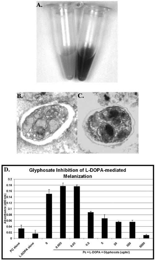
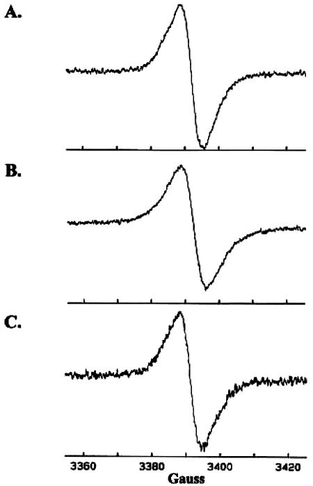
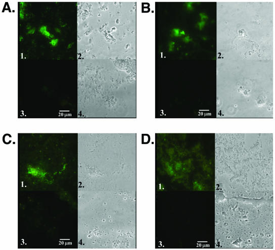
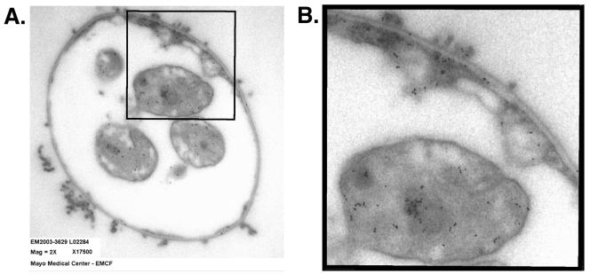
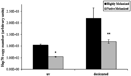
Pneumocystis continues to represent an important opportunistic fungal pathogen of those with compromised immunity. Thus, it is crucial to identify factors that affect its viability and pathogenicity. We previously reported the first identification of melanins in Pneumocystis. In the present study, we sought to further characterize these components and define the function for these melanins. Melanins extracted from Pneumocystis and melanized Pneumocystis cells were analyzed by electron spin resonance spectroscopy, revealing spectra consistent with melanins from other fungi. Immunofluorescence assays using anti-melanin monoclonal antibodies showed that melanins are widely present across Pneumocystis host species, including mouse-, ferret-, and human-derived Pneumocystis organisms, as well as Pneumocystis carinii derived from rat. Using immunoelectron microscopy, melanins were found to localize to the cell wall and cytoplasm of P. carinii cysts, as well as to intracystic bodies within mature cysts. Next, the role of melanins on the maintenance of Pneumocystis viability was determined by using quantitative reverse transcription-PCR measurement of the heat shock protein mRNA under adverse environmental conditions. Using a new method to promote the melanization of Pneumocystis, we observed that strongly melanized Pneumocystis retained viability to a greater degree when exposed to UV irradiation or desiccation compared to less-pigmented organisms. These studies support our previous identification of Pneumocystis melanins across the genus, further characterize these Pneumocystis components, and demonstrate that melanins protect Pneumocystis from environmental stressors.
Figures
Similar articles
-
Evidence for Proinflammatory β-1,6 Glucans in the Pneumocystis carinii Cell Wall.Infect Immun. 2015 Jul;83(7):2816-26. doi: 10.1128/IAI.00196-15. Epub 2015 Apr 27. Infect Immun. 2015. PMID: 25916991 Free PMC article.
-
Evidence for a melanin cell wall component in Pneumocystis carinii.Infect Immun. 2003 Sep;71(9):5360-3. doi: 10.1128/IAI.71.9.5360-5363.2003. Infect Immun. 2003. PMID: 12933884 Free PMC article.
-
[Presence of Pneumocystis carinii and Pneumocystis wakefieldiae DNA in the extrapulmonary tissues of immunocompromised laboratory rats].Wiad Parazytol. 2009;55(2):167-71. Wiad Parazytol. 2009. PMID: 19670532 Polish.
-
Pneumocystis carinii: an update.Ultrastruct Pathol. 2003 Mar-Apr;27(2):115-22. doi: 10.1080/01913120309928. Ultrastruct Pathol. 2003. PMID: 12746203 Review.
-
Pathobiology of Pneumocystis pneumonia: life cycle, cell wall and cell signal transduction.FEMS Yeast Res. 2015 Sep;15(6):fov046. doi: 10.1093/femsyr/fov046. Epub 2015 Jun 12. FEMS Yeast Res. 2015. PMID: 26071598 Review.
Cited by
-
Retracing the evolution of Pneumocystis species, with a focus on the human pathogen Pneumocystis jirovecii.Microbiol Mol Biol Rev. 2024 Jun 27;88(2):e0020222. doi: 10.1128/mmbr.00202-22. Epub 2024 Apr 8. Microbiol Mol Biol Rev. 2024. PMID: 38587383 Free PMC article. Review.
-
A Molecular Window into the Biology and Epidemiology of Pneumocystis spp.Clin Microbiol Rev. 2018 Jun 13;31(3):e00009-18. doi: 10.1128/CMR.00009-18. Print 2018 Jul. Clin Microbiol Rev. 2018. PMID: 29899010 Free PMC article. Review.
-
Parallels in fungal pathogenesis on plant and animal hosts.Eukaryot Cell. 2006 Dec;5(12):1941-9. doi: 10.1128/EC.00277-06. Epub 2006 Oct 13. Eukaryot Cell. 2006. PMID: 17041185 Free PMC article. Review. No abstract available.
-
Evidence for Proinflammatory β-1,6 Glucans in the Pneumocystis carinii Cell Wall.Infect Immun. 2015 Jul;83(7):2816-26. doi: 10.1128/IAI.00196-15. Epub 2015 Apr 27. Infect Immun. 2015. PMID: 25916991 Free PMC article.
-
Impact of melanin on microbial virulence and clinical resistance to antimicrobial compounds.Antimicrob Agents Chemother. 2006 Nov;50(11):3519-28. doi: 10.1128/AAC.00545-06. Antimicrob Agents Chemother. 2006. PMID: 17065617 Free PMC article. Review. No abstract available.
References
-
- Bell, A. A., and M. H. Wheeler. 1986. Biosynthesis and functions of fungal melanins. Annu. Rev. Phytopathol. 24:411-451.
-
- Bernard, M., and J. P. Latge. 2001. Aspergillus fumigatus cell wall: composition and biosynthesis. Med. Mycol. 39:9-17. - PubMed
-
- Butler, M. J., and A. W. Day. 1998. Fungal melanins: a review. Can. J. Microbiol. 44:1115-1136.
-
- Desai, J. D., and V. V. Modi. 1977. Growth, glucose metabolism and melanin formation in biotin-deficient Aspergillus nidulans. Folia Microbiol. 22:55-60. - PubMed
Publication types
MeSH terms
Substances
Grants and funding
LinkOut - more resources
Full Text Sources
