. 2006 Jun;5(6):981-90.
doi: 10.1128/EC.00052-06.

Investigation of the mechanism of meiotic DNA cleavage by VMA1-derived endonuclease uncovers a meiotic alteration in chromatin structure around the target site

Affiliations

Investigation of the mechanism of meiotic DNA cleavage by VMA1-derived endonuclease uncovers a meiotic alteration in chromatin structure around the target site

Tomoyuki Fukuda et al. Eukaryot Cell. 2006 Jun.

Abstract

VMA1-derived endonuclease (VDE), a homing endonuclease in Saccharomyces cerevisiae, is encoded by the mobile intein-coding sequence within the nuclear VMA1 gene. VDE recognizes and cleaves DNA at the 31-bp VDE recognition sequence (VRS) in the VMA1 gene lacking the intein-coding sequence during meiosis to insert a copy of the intein-coding sequence at the cleaved site. The mechanism underlying the meiosis specificity of VMA1 intein-coding sequence homing remains unclear. We studied various factors that might influence the cleavage activity in vivo and found that VDE binding to the VRS can be detected only when DNA cleavage by VDE takes place, implying that meiosis-specific DNA cleavage is regulated by the accessibility of VDE to its target site. As a possible candidate for the determinant of this accessibility, we analyzed chromatin structure around the VRS and revealed that local chromatin structure near the VRS is altered during meiosis. Although the meiotic chromatin alteration exhibits correlations with DNA binding and cleavage by VDE at the VMA1 locus, such a chromatin alteration is not necessarily observed when the VRS is embedded in ectopic gene loci. This suggests that nucleosome positioning or occupancy around the VRS by itself is not the sole mechanism for the regulation of meiosis-specific DNA cleavage by VDE and that other mechanisms are involved in the regulation.


Figures

FIG. 1.
FIG. 1.
Examination of endonuclease activity and effects of nuclear localization or meiotic progression on meiosis-specific homing. (A) Endonuclease activity of immunoprecipitated VDE. Cells expressing Flag-tagged VDE were subjected to synchronous sporulation and sampled at various time points in SPM. Using an anti-Flag antibody, VDE was immunoprecipitated and assayed by cleavage of the 3.3-kb linearized DNA containing the VRS that produces 2.2- and 1.1-kb fragments after digestion at the VRS. Substrates were incubated for 0, 15, 30, and 60 min at 30°C. (B) Physical detection of homing by Southern blotting. VMA1-207/VMA1 heterozygous diploid cells ectopically expressing no VDE, wild-type VDE, or NLS-fused VDE were subjected to synchronous sporulation. Since the VMA1-207 allele contains an intein-coding sequence encoding inactive VDE (R90A), only VDE that is ectopically expressed can produce DSBs and the cleaved recipient is repaired using a VMA1-207 donor as a template, resulting in the production of a VMA1+ homing product. DNA was isolated from cells at the indicated times after incubation in SPM and subjected to Southern analysis after BamHI digestion. The BamHI site polymorphism makes it possible to distinguish VMA1+ homing products from VMA1-207 donors. (C) Physical analysis of homing in mutants deficient in progression of meiotic program. Southern analysis was performed on the VMA1+/VMA1 heterozygous diploid cells in which meiotic entry, DNA replication, or nuclear divisions were blocked by the introduction of each mutation. (D) VDE localization in meiotic mutants at 4 h in SPM. The upper panels show localization of VDE, and the lower panels show the nuclei. DAPI, 4′,6′-diamidino-2-phenylindole.
FIG. 2.
FIG. 2.
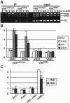
Detection of VDE binding to the VRS. (A) Diploid cells homozygous for VMA1- expressing VDE or DNA-binding-deficient VDE (R90A) were sampled at the indicated times during meiosis. Cells were treated with formaldhyde, and then the cell extract was prepared, sonicated to shear the chromatin, and immunoprecipitated with anti-VDE antibody. DNA from input samples and in the immunoprecipitates (IP) was amplified by multiplex PCR with primers for the 3′ region adjacent to the VRS (VRS3), the 5′ region adjacent to the VRS (VRS5), and the telomeric region of chromosome VI (TEL). (B) The ratios of the VRS signals to the TEL signal were normalized to those of the input samples at each time point. Error bars denote the standard deviations among three independent experiments. (C) A ChIP assay with anti-VDE antibody was performed for the indicated mutants. The signals obtained for the 4.5-h samples are shown as in panel B.
FIG. 3.
FIG. 3.
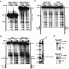
Chromatin configuration at the VMA1 locus during meiosis. (A) Chromatin was prepared in diploid cells homozygous for VMA1 at various times of incubation in SPM, treated with 0, 5, 10, or 20 U/ml of MNase, and redigested with BamHI and SphI. Naked DNA was also treated with 0.2, 0.4, or 0.8 U/ml of MNase. MNase-sensitive sites were detected by indirect end-labeling using a probe for the sequence adjacent to the BamHI site. The vertical gray arrow indicates the position of the coding region of the VMA1 locus. The arrowheads show MNase-sensitive sites near the VRS that became prominent during meiosis. Numbers indicate the positions in nucleotides relative to the VDE cutting site that is shown by the horizontal arrow. Meiotic progression was monitored by DNA content, which was analyzed by a fluorescence-activated cell sorter (B), and by nuclear division, which was analyzed by microscopy of 4′,6′-diamidino-2-phenylindole-stained cells (C).
FIG. 4.
FIG. 4.
Chromatin alteration during meiosis occurs independently of VDE expression and is dependent on meiotic progression. Results of chromatin analysis of VMA1 homozygous cells expressing wild-type VDE (A), endonuclease-defective VDE (D326V) (B), and VDE defective in DNA binding (R90A) (B) are shown. Samples were treated with 0, 5, or 10 U/ml of MNase. Indirect end-labeling was performed, and MNase-sensitive sites were analyzed as described in the legend to Fig. 3. The arrowheads indicate the chromatin alteration site. (C) Chromatin structure of the haploid cell in SPM. Chromatin was prepared in VMA1 haploid cells sampled at the indicated times of incubation in SPM and treated with 0, 5, or 10 U/ml of MNase. The sir2Δ mutation permits haploid cells to enter the meiotic program. (C) Chromatin structure around the VRS in mutant cells defective in meiotic progression. Chromatin was prepared from VMA1 homozygous diploid cells after 1.5 and 4.5 h of incubation in SPM and subjected to indirect end-labeling.
FIG. 5.
FIG. 5.
(A) Chromatin structure around the VRS in diploid cells homozygous for VMA1 or VMA1+. In the VMA1+ allele, the VRS is interrupted by the insertion of an intein-coding sequence in the middle of the sequence. Samples were treated with 0, 5, or 10 U/ml of MNase. (B) Chromatin configuration at the ADE2 gene locus. Mitotic (0 h) and meiotic (4 h) chromatins were prepared from diploid cells homozygous for ADE2 or VRS-inserted ADE2 (designated ADE2::VRS), treated with 0, 5, 10, or 20 U/ml of MNase, and redigested with AseI. MNase-sensitive sites were detected by indirect end-labeling with a probe for the sequence adjacent to the AseI site. The vertical gray arrow indicates the position of the coding region for the ADE2 locus. The arrowheads show MNase-sensitive sites that became prominent in ADE2::VRS during meiosis. The right panel shows the positions of the VDE cutting site (horizontal arrow) and the chromatin alteration site (arrowhead). Numbers indicate the positions in nucleotides relative to the VDE cutting site. (C) Chromatin configuration at the MCM4 gene locus. Mitotic (0 h) and meiotic (4 h) chromatins were prepared from diploid cells homozygous for MCM4 or VRS-inserted MCM4 (designated MCM4::VRS) and then analyzed by end-labeling after treatment with 0, 5, or 10 U/ml of MNase. (D) Physical detection of the VDE-mediated DSB by Southern blotting. VMA1+ diploid cells homozygous for ADE2::VRS or MCM4::VRS were subjected to synchronous sporulation. DNA was isolated from cells at the indicated times after incubation in SPM and subjected to Southern analysis.

Similar articles

Cited by

References

    1. Belfort, M., and R. J. Roberts. 1997. Homing endonucleases: keeping the house in order. Nucleic Acids Res. 25:3379-3388. - PMC - PubMed
    1. Burt, A., and V. Koufopanou. 2004. Homing endonuclease genes: the rise and fall and rise again of a selfish element. Curr. Opin. Genet. Dev. 14:609-615. - PubMed
    1. Chevalier, B. S., and B. L. Stoddard. 2001. Homing endonucleases: structural and functional insight into the catalysts of intron/intein mobility. Nucleic Acids Res. 29:3757-3774. - PMC - PubMed
    1. Chu, S., and I. Herskowitz. 1998. Gametogenesis in yeast is regulated by a transcriptional cascade dependent on Ndt80. Mol. Cell 1:685-696. - PubMed
    1. Church, G. M., and W. Gilbert. 1984. Genomic sequencing. Proc. Natl. Acad. Sci. USA 81:1991-1995. - PMC - PubMed

Publication types

MeSH terms