. 2006 Jun 7;25(11):2615-22.
doi: 10.1038/sj.emboj.7601167. Epub 2006 Jun 1.

Ser18 and 23 phosphorylation is required for p53-dependent apoptosis and tumor suppression

Affiliations

Ser18 and 23 phosphorylation is required for p53-dependent apoptosis and tumor suppression

Connie Chao et al. EMBO J. .

Abstract

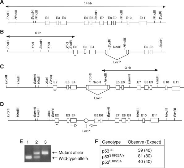
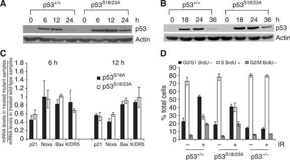
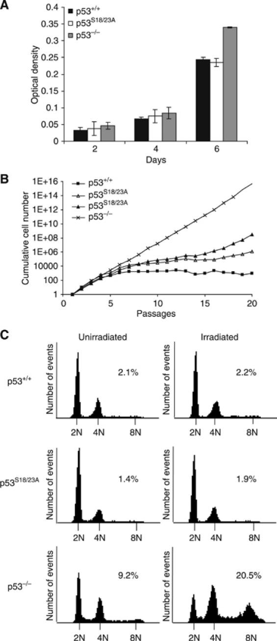
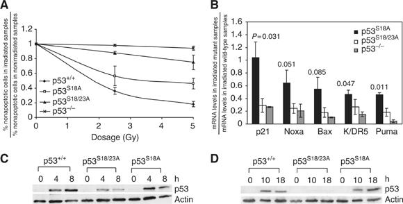
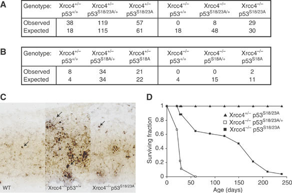
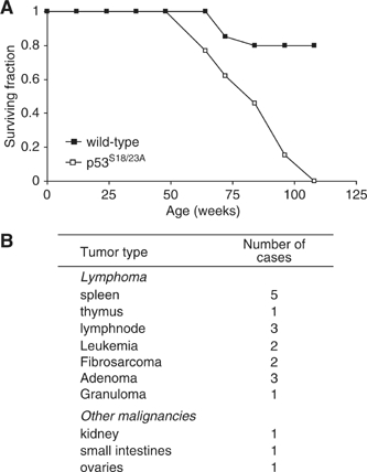
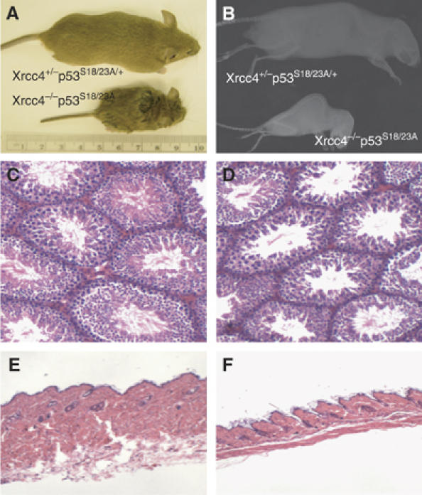
Mouse p53 is phosphorylated at Ser18 and Ser23 after DNA damage. To determine whether these two phosphorylation events have synergistic functions in activating p53 responses, we simultaneously introduced Ser18/23 to Ala mutations into the endogenous p53 locus in mice. While partial defects in apoptosis are observed in p53S18A and p53S23A thymocytes exposed to IR, p53-dependent apoptosis is essentially abolished in p53S18/23A thymocytes, indicating that these two events have critical and synergistic roles in activating p53-dependent apoptosis. In addition, p53S18/23A, but not p53S18A, could completely rescue embryonic lethality of Xrcc4(-/-) mice that is caused by massive p53-dependent neuronal apoptosis. However, certain p53-dependent functions, including G1/S checkpoint and cellular senescence, are partially retained in p53(S18/23A) cells. While p53(S18A) mice are not cancer prone, p53S18/23A mice developed a spectrum of malignancies distinct from p53S23A and p53(-/-) mice. Interestingly, Xrcc4(-/-)p53S18/23A mice fail to develop tumors like the pro-B cell lymphomas uniformly developed in Xrcc4(-/-) p53(-/-) animals, but exhibit developmental defects typical of accelerated ageing. Therefore, Ser18 and Ser23 phosphorylation is important for p53-dependent suppression of tumorigenesis in certain physiological context.


Figures

Figure 1
Figure 1
Generation of p53S18/23A knockin mice. (A) The endogenous mouse p53 locus. (B) The targeting construct. The PGK-NeoR cassette flanked by two loxP sites was inserted into intron 4. S18/23A mutations are indicated by asterisks. (C) The targeted p53 locus following homologous recombination between the endogenous p53 allele and targeting vector. (D) Knockin p53 allele following LoxP/Cre-mediated deletion of the PGK-NeoR gene. (E) PCR analysis of the genomic DNA isolated from the tails of WT (lane 1), p53S18/23A/+ (lane 2) and p53S18/23A mice (lane 3). The PCR products depicting the WT and knockin mutant allele are indicated to the right. The primers are indicated by arrowheads. (F) The number of offsprings in various genotypes derived from the breeding of p53S18/23A/+ mice. The expected number based on the Mendelian ratio is also shown.
Figure 2
Figure 2
p53 stabilization and activity in p53S18/23A MEFs after DNA damage. Protein levels of p53 in the WT and p53S18/23A MEFs at various time points after exposure to 0.25 μM doxorubicin (A) or 60 J/M2 UVC (B). Time points and genotypes are indicated on the top. p53 and actin are indicated on the right. (C) Real-time PCR analysis of the p53-dependent transcription of p21, Noxa, Bax and K/DR5 at 6 and 12 h after doxorubicin treatment. The ratio of the mRNA levels in the untreated p53S18A or p53S18/23A MEFs versus those in treated WT MEFs is shown. Mean values from three independent experiments are shown with standard deviation. (D) Cell cycle G1/S arrest in WT, p53S18/23A and p53−/− MEFs after 20 Gy of IR. The genotypes are indicated at the bottom.
Figure 3
Figure 3
Cellular proliferation and polyploidy of p53S18/23A cells. (A) Proliferation of thymocytes derived from WT, p53S18/23A and p53−/− mice. Thymocytes were activated with 5 ng/ml PMA, 500 ng/ml ionomycin, and proliferation was measured after 2, 4 or 6 days of stimulation. Mean values from triplicate wells are shown with error bars. (B) 3T3 proliferation assay of WT, p53S18/23A and p53−/− MEFs. MEFs were serially passaged with a plating density of 3 × 10⁵ cells per 6 cm plate and counted once every 3 days up to 20 passages. Cumulative cell numbers, based on the average of duplicate plates for each passage, are shown. Data from two independent p53S18/23A MEFs are presented. (C) Polyploidy in WT, p53S18/23A and p53−/− MEFs before (left panels) and 24 h after IR (right panels). Genotypes are indicated to the left. Histograms show DNA content on X-axis versus cell number on Y-axis. The peaks representing 2N, 4N and 8N cells are indicated. The percentage of total cells with over 8N DNA content is shown.
Figure 4
Figure 4
p53 stabilization and activity in p53S18/23A thymocytes after IR. (A) p53-dependent apotosis of WT, p53−/−, p53S18A and p53S18/23A thymocytes 24 h after exposure to 2.5 Gy and 5 Gy IR. Mean values from three independent experiments are shown with standard deviation. (B) p53-dependent transcription of its target genes in p53S18A, p53S18/23A and p53−/− thymocytes as compared to that in WT thymocytes 18 h after 5 Gy IR. Mean values from two independent experiments are shown with error bars. P-values between the levels of reduction in p53S18/23A thymocytes and p53S18A thymocytes are given. Protein levels of p53 in WT, p53S18/23A and p53S18A thymocytes at earlier (C) or later time points (D) after 5 Gy IR. Genotypes and time points are indicated on the top. p53 and actin are indicated on the right.
Figure 5
Figure 5
p53S18/23A mutation completely rescues the embryonic lethality of Xrcc4−/− mice. (A) The number of offspring in various genotypes derived from the following breeding: Xrcc4+/−p53S18/23A/+ × Xrcc4+/−p53S18/23A/+ or Xrcc4+/−p53S18/23A × Xrcc4+/−p53S18/23A. The expected number based on the Mendelian ratio is also shown. (B) The number of offsprings in various genotypes derived from either Xrcc4+/−p53S18A/+ × Xrcc4+/−p53S18A/+ or Xrcc4+/−p53S18A × Xrcc4+/−p53S18A breeding. (C) p53-dependent apoptosis in the embryonic cerebral cortex as shown by ISEL+. Saggital sections from E12.5 embryos were shown. The apoptotic cells are indicated by arrowheads. (D) The surviving percentage of Xrcc4+/−p53S18/23A (n=16), Xrcc4−/−p53S18/23A/+ (n=10) and Xrcc4−/−p53S18/23A (n=34) mice at various times after birth. N represents the number of mice monitored. P-value is 0.0004 for the comparison between Xrcc4−/−p53S18/23A/+ and Xrcc4+/−p53S18/23A mice and 0.001 for the comparison between Xrcc4−/−p53S18/23A and Xrcc4+/−p53S18/23A mice.
Figure 6
Figure 6
Aging p53S18/23A mice are prone to spontaneous cancer. (A) Survival curve of 20 pairs of WT and p53S18/23A mice. P-value is 0.04 for the comparison of the survival rate of p53S18/23A mice and WT controls. Animals were monitored for spontaneous tumorigenesis over the course of 2 years. (B) The spectrum of tumors developed in p53S18/23A mice.
Figure 7
Figure 7
Xrcc4−/−p53S18/23A mice show accelerated aging phenotype. (A) Picture of 8-week-old male Xrcc4+/−p53S18/23A/+ and Xrcc4−/−p53S18/23A littermates. Histological analysis of testes (C, D) and skin (E, F) of the 8-week-old Xrcc4+/−p53S18/23A/+ (C, E) and Xrcc4−/−p53S18/23A (D, F) littermates. (B) X-ray image showing the spine curvature of 17-week-old Xrcc4+/−p53S18/23A/+ and Xrcc4−/−p53S18/23A littermates.

References

    1. Bischoff F, Yim S, Pathak S, Grant G, Siciliano M, Giovanella B, Strong L, Tainsky M (1990) Spontaneous abnormalities in normal fibroblasts from patients with Li-Fraumeni cancer syndrome: aneuploidy and immortalization. Cancer Res 50: 7979–7984 - PubMed
    1. Blaschke JAW, Chun J (1998) Programmed cell death is a universal feature of embryonic and postnatal neuroproliferative regions throughout the central nervous system. J Comp Neurol 396: 39–50 - PubMed
    1. Blaschke A, Staley K, Chun J (1996) Widespread programmed cell death in proliferative and postmitotic regions of the fetal cerebral cortex. Development 122: 1165–1174 - PubMed
    1. Borges HL, Chao C, Xu Y, Linden R, Wang JY (2004) Radiation-induced apoptosis in developing mouse retina exhibits dose-dependent requirement for ATM phosphorylation of p53. Cell Death Differ 11: 494–502 - PubMed
    1. Bouffler S, Kemp C, Balmain A, Cox R (1995) Spontaneous and ionizing radiation-induced chromosomal abnormalities in p53-deficient mice. Cancer Res 55: 3883–3889 - PubMed

Publication types

MeSH terms