. 2006 Jun 15;49(12):3553-62.
doi: 10.1021/jm060092o.

Finding a needle in a haystack: development of a combinatorial virtual screening approach for identifying high specificity heparin/heparan sulfate sequence(s)

Affiliations

Finding a needle in a haystack: development of a combinatorial virtual screening approach for identifying high specificity heparin/heparan sulfate sequence(s)

Arjun Raghuraman et al. J Med Chem. .

Abstract

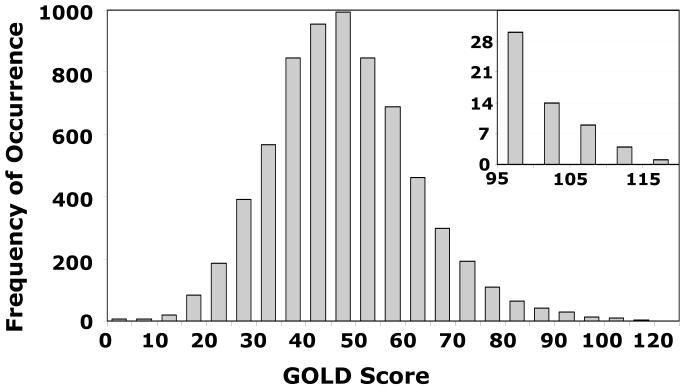
We describe a combinatorial virtual screening approach for predicting high specificity heparin/heparan sulfate sequences using the well-studied antithrombin-heparin interaction as a test case. Heparan sulfate hexasaccharides were simulated in the 'average backbone' conformation, wherein the inter-glycosidic bond angles were held constant at the mean of the known solution values, irrespective of their sequence. Molecular docking utilized GOLD with restrained inter-glycosidic torsions and intra-ring conformations, but flexible substituents at the 2-, 3-, and 6-positions and explicit incorporation of conformational variability of the iduronate residues. The approach reproduces the binding geometry of the sequence-specific heparin pentasaccharide to within 2.5 A. Screening of a combinatorial virtual library of 6,859 heparin hexasaccharides using a dual filter strategy, in which predicted antithrombin affinity was the first filter and self-consistency of docking was the second, resulted in only 10 sequences. Of these, nine were found to bind antithrombin in a manner identical to the natural pentasaccharide, while a novel hexasaccharide bound the inhibitor in a unique but dramatically different geometry and orientation. This work presents the first approach on combinatorial library screening for heparin/heparan sulfate GAGs to determine high specificity sequences and opens up huge opportunities to investigate numerous other physiologically relevant GAG-protein interactions.


Figures

Figure 1
Figure 1. Structure of natural pentasaccharide H5 and its derivative H5CRYS extracted from crystal structure study of Li et al and used in docking studies
D, E, F, G, and H refer to historical label of residues from the non-reducing end. Sulfate groups in H5 highlighted as filled ovals formula image 
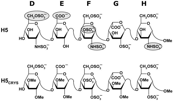
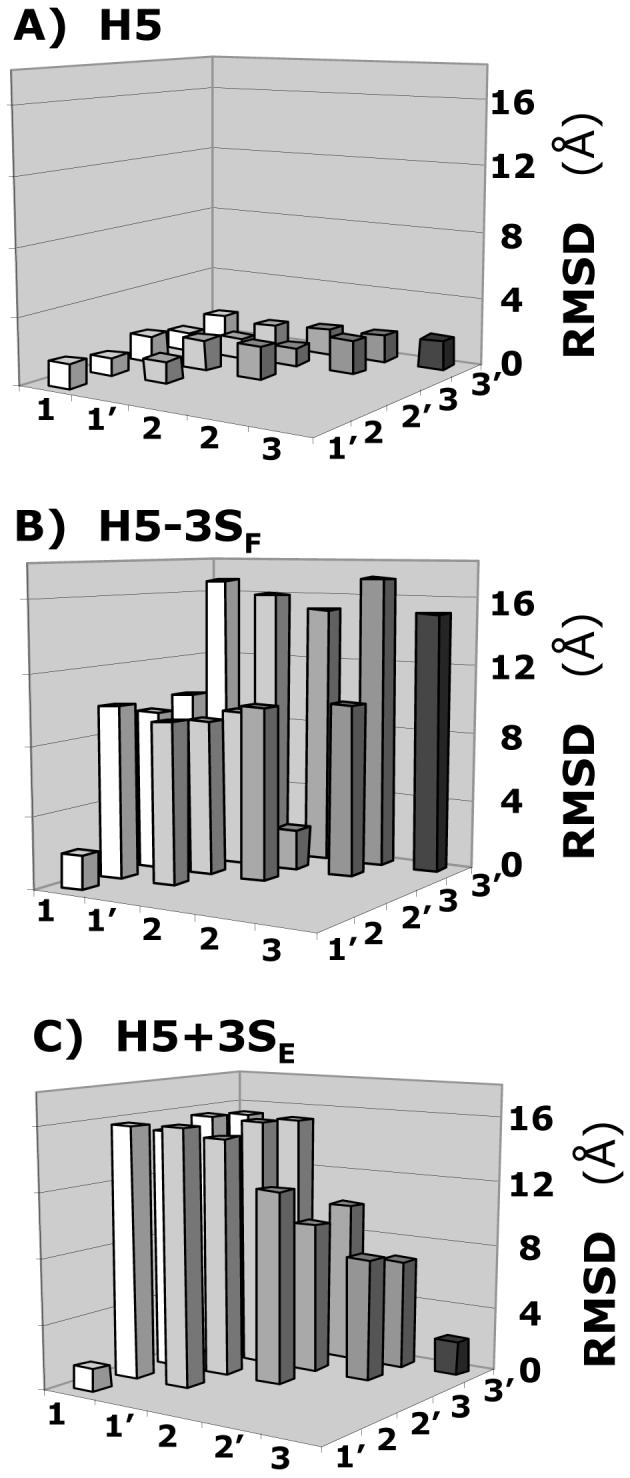
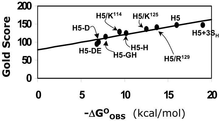
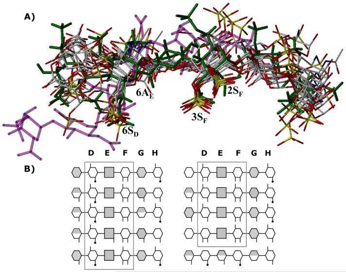
are critical for high-affinity interaction with antithrombin.
Figure 2
Figure 2. Structures of H5 and its truncated variants studied for docking
D, E, F, G, and H refer to residue labels. Variations in structure (V, W, X, Y, Z, and Z’) to give variants with either one more sulfate group (H5+3SH, H5+3SE and H5+3SG) or one less sulfate group (H5-6SD, H5-3SF, H5-2SH) are highlighted as filled ovals formula image
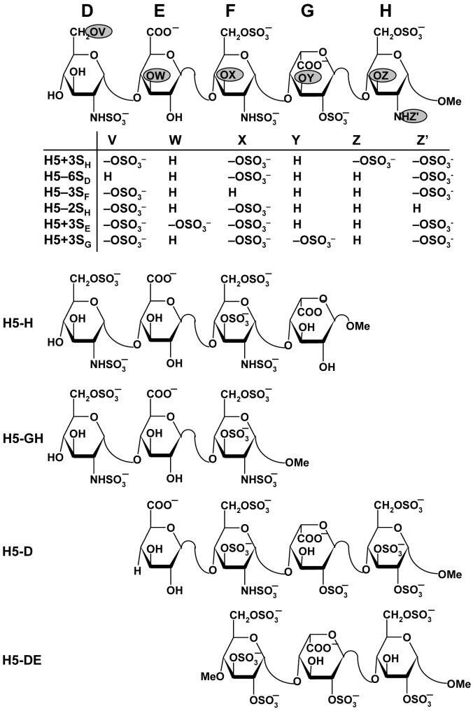
. Truncated pentasaccharides H5-H, H5-GH, H5-D, and H5-DE refer to one or two residue deletion variants from the either end.
Figure 3
Figure 3. Disaccharide sequences used to build virtual library of 6,859 hexasaccharides
Although theoretically 48 disaccharides are possible, 23 have been experimentally observed. Based on the sequence of H5, our library included GlcAp sequences with GlcNp3S and IdoAp sequences without GlcNp3S, with IdoAp in either 2SO or 1C4 conformation. These variations made up 19 disaccharide sequences to produce 19×19×19 = 6,859 hexasaccharides.
Figure 4
Figure 4. Comparison of GOLD predicted binding geometry of natural pentasaccharide H5 having ‘average backbone’ with that of H5CRYS determined in the crystal structure
H5 with average inter-glycosidic bond parameters (‘the average backbone’), was docked onto antithrombin. Structurally H5 differs from H5CRYS (see Fig. 1). An overlay of 6 solutions from three independent docking runs shows high consistency in the predicted binding geometry, which matches the crystal structure geometry with an RMSD less than 2.5 Å. Structure in green is the crystal structure geometry, while those in atom-type color (red, yellow, grey and blue) are 6 docking solutions. Note the identical orientation of key groups, 2- and 3-OSO3- of residue F (2SF and 3SF), 6-COO- of residue E (6AE) and 6-OSO3- of residue D (6SD). Helices A, D and P of antithrombin (in ribbon diagram) are indicated as hA, hD and hP, while D, E, F, G, and H labels correspond to residues of the pentasaccharide. K114, K125 and R129 are shown in ball and stick representation. See text for details.
Figure 5
Figure 5. Specificity of GAG sequence for antithrombin
Variation in the RMSD between six solutions (1, 1′, 2, 2′, 3, and 3′) obtained from three independent docking experiments of heparin pentasaccharide H5 variants, containing either one additional sulfate group at 3-position of residue E or lacking a sulfate at the 3-position of residue F, binding to antithrombin. Whereas for H5 (A) the RMSD was less than 2.0 Å between each solution, for H5-3SF (B) and H5+3SE (C), a majority of RMSD values were much greater (5-17 Å). Other pentasaccharide variants were also studied. See text for details.
Figure 6
Figure 6. Correlation of GOLD score with antithrombin binding affinity
Modified GOLD score (See Methods) determined following 100,000 iterations and multiple docking runs linearly increases with the observed antithrombin binding affinity under physiological conditions. Only those antithrombin complexes for which binding affinities were measured under essentially equivalent conditions are used for analysis. Labels refer to pentasaccharide or its variant, identified in Figures 1 and 2, interaction with wild-type antithrombin. H5/K114, H5/K125 and H5/R129 refer to natural pentasaccharide H5 interaction with mutant antithrombins.- Solid line represents linear regression line with slope of 4.2 GOLD score per kcal/mol and an intercept of 78.9 GOLD score. See text for details.
Figure 7
Figure 7. Histogram of number of HS hexasaccharide sequences for every 5 unit change in GOLD score
Modified GOLD score was calculated for all 6,859 hexasaccharides docked onto antithrombin following the first phase of combinatorial library screening. Inset shows an expanded portion of the histogram in the range 95-120 GOLD score. See text for details.
Figure 8
Figure 8. Finding a needle in a haystack
A) An overlay of final 10 hexasaccharide sequences obtained after second phase of combinatorial library screening. Structure in green is the H5CRYS, while those in atom-type color are 9 sequences with nearly identical binding orientation and geometry. Sequence in purple color was found to bind antithrombin reproducibly with high specificity and affinity but dramatically different orientation. Labels 2SF, 3SF, 6AE and 6SD represent sulfate or carboxylate groups at the 2- and 3-position of residue F, 6-position of residue E and 6-positon of residue D. B) Symbolic representation of the high-affinity, high-specificity hexasaccharide structures shown above. The hexasaccharide library sequence runs {UAp(1→4)GlcNp(1→4)}3, where UA is either IdoAp (shaded hexagon) or GlcAp (shaded square). Sulfated substitution at 2-, 3- or 6-positions of either UAp or GlcNp is indicated with a line (—), while acetate substitution at the 2-positon of GlcNp is indicated with a line-dot (—·). Iduronic acid residues in 2SO conformation are shown as fully shaded hexagons, while those in 1C4 conformation are shown as half-filled hexagons. See text for details. See Supplementary Information for a MOL2 file of all the combinatorial sequences.

References

    1. Pike RN, Buckle AM, Le Bonniec BF, Church FC. Control of the coagulation system by serpins. Getting by with a little help from glycosaminoglycans. FEBS J. 2005;272:4842–4851. - PubMed
    1. Desai UR. Antithrombin activation and designing novel heparin mimics. In: Garg HG, Linhardt RJ, Hales CA, editors. Chemistry and Biology of Heparin and Heparan Sulfate. Elsevier; NY: 2005. pp. 483–513.
    1. Presta M, Dell’Era P, Mitola S, Moroni E, Ronca R, Rusnati M. Fibroblast growth factor/fibroblast growth factor receptor system in angiogenesis. Cytokine Growth Factor Rev. 2005;16:159–178. - PubMed
    1. Rops AL, van der Vlag J, Lensen JF, Wijnhoven TJ, van den Heuvel LP, van Kuppevelt TH, Berden JH. Heparan sulfate proteoglycans in glomerular inflammation. Kidney Int. 2004;65:768–785. - PubMed
    1. Kelton JG. The pathophysiology of heparin-induced thrombocytopenia: biological basis for treatment. Chest. 2005;127:9S–20S. - PubMed

Publication types