. 2006 Nov;291(5):C946-56.
doi: 10.1152/ajpcell.00014.2006. Epub 2006 Jun 7.

Effects of alpha1D-adrenergic receptors on shedding of biologically active EGF in freshly isolated lacrimal gland epithelial cells

Affiliations

Effects of alpha1D-adrenergic receptors on shedding of biologically active EGF in freshly isolated lacrimal gland epithelial cells

LiLi Chen et al. Am J Physiol Cell Physiol. 2006 Nov.

Abstract

Transactivation of EGF receptors by G protein-coupled receptors is a well-known phenomenon. This process involves the ectodomain shedding of growth factors in the EGF family by matrix metalloproteinases. However, many of these studies employ transformed and/or cultured cells that overexpress labeled growth factors. In addition, few studies have shown that EGF itself is the growth factor that is shed and is responsible for transactivation of the EGF receptor. In this study, we show that freshly isolated, nontransformed lacrimal gland acini express two of the three known alpha(1)-adrenergic receptors (ARs), namely, alpha(1B)- and alpha(1D)-ARs. Alpha(1D)-ARs mediate phenylephrine (an alpha(1)-adrenergic agonist)-induced protein secretion and activation of p42/p44 MAPK, because the alpha(1D)-AR inhibitor BMY-7378, but not the alpha(1A)-AR inhibitor 5-methylurapidil, inhibits these processes. Activation of p42/p44 MAPK occurs through transactivation of the EGF receptor, which is inhibited by the matrix metalloproteinase ADAM17 inhibitor TAPI-1. In addition, phenylephrine caused the shedding of EGF from freshly isolated acini into the buffer. Incubation of freshly isolated cells with conditioned buffer from cells treated with phenylephrine resulted in activation of the EGF receptor and p42/p44 MAPK. The EGF receptor inhibitor AG1478 and an EGF-neutralizing antibody blocked this activation of p42/p44 MAPK. We conclude that in freshly isolated lacrimal gland acini, alpha(1)-adrenergic agonists activate the alpha(1D)-AR to stimulate protein secretion and the ectodomain shedding of EGF to transactivate the EGF receptor, potentially via ADAM17, which activates p42/p44 MAPK to negatively modulate protein secretion.


Figures
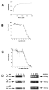
Figure 1
Figure 1. Binding, Competition and RT-PCR Studies
Saturation studies were performed by incubating lacrimal gland membranes with increasing concentrations of 125I-HEAT in the presence or absence of 0.1mM phentolamine. Specific binding is shown for a representative curve (A). Bmax= 0.206 ± 0.033 pmoles/mg membrane protein; KD = 107.8 ± 10.7 pM from 3 independent experiments performed in triplicate. Competition studies were performed using the α1A-adrenergic receptor selective inhibitor 5-methylurapidil (5-MU) (B) or the α1D-adrenergic receptor selective inhibitor BMY7378 (C) from 10pM to 1mM in the presence of 90pM 125I-HEAT. Various symbols present data from individual experiments (n=4), performed in duplicate. (D) RT-PCR was performed on lacrimal gland acini and brain mRNA using primers specific to α1A-, α1B-, α1D-adrenergic receptors or G3PDH. Each lane (1, 2, and 3) corresponds to an individual animal. Lane 4 corresponds to RT-PCR performed with each primer using brain mRNA. NC – Negative Control.
Figure 2
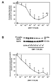
Figure 2. Effect of Inhibition of α1D-Adrenergic Receptors on Lacrimal Gland Functions
Lacrimal gland acini were preincubated for 20 minutes with the specific α1D-adrenergic receptor inhibitor BMY-7378 (10−11 –10−5 M) prior to addition of the α1-adrenergic agonist phenylephrine (10−4 M) and (A) peroxidase secretion was measured after 20 minutes. Data are mean ± SEM of percent of response from 3–6 independent experiments. (B) p42/p44 MAPK was measured after 5 minutes. Data are mean ± SEM of percent of response from 4–8 independent experiments. The blot shown in B is a representative blot. * indicates statistical significance from no BMY-7378; # indicates statistical significance from 10−5 M BMY-7378.
Figure 3
Figure 3. Effect of Inhibition of Matrix Metalloproteinases on Lacrimal Gland Functions
Lacrimal gland acini were preincubated for 20 minutes with either GM6001, its negative control (10−6 M, A) or ADAM-17 inhibitor TAPI-1 (10−4 –10−6 M, B) prior to addition of the α1-adrenergic agonist phenylephrine (10−4 M) and peroxidase secretion was measured after 20 minutes. Data are mean ± SEM from 6 independent experiments. Acini were preincubated for 20 minutes with either GM6001, its negative control (10−6 M, E) or ADAM-17 inhibitor TAPI-1 (10−4 –10−6 M, F) prior to addition of the α1-adrenergic agonist phenylephrine (10−4 M) and p42/p44 MAPK was measured after 5 minutes. Data are mean ± SEM from 6 independent experiments. Blots shown in C and D are representative experiments. * indicates statistical significance from agonist alone.
Figure 4
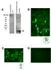
Figure 4. Presence of EGF in the Lacrimal Gland
(A) Proteins from either lacrimal gland homogenate or acini were separated on 4–20% polyacrylamide gel and EGF was detected in whole lacrimal gland, isolated lacrimal gland acini, commercially available rat EGF, and whole submandibular gland using an anti-EGF antibody. Arrow indicates the 6 kDa EGF form of EGF. (B) Localization of EGF was determined by immunofluorescence techniques in lacrimal gland. * indicate the presence of EGF in lacrimal gland ducts; arrows indicate the presence of EGF in the apical and lateral membranes of lacrimal gland acinar cells. Inset shows presence of EGF in apical and lateral membranes of lacrimal gland acinar membranes. A schematic diagram of the structure of a lacrimal gland acinus is below with the apical membrane highlighted in red and with the red arrowhead. Green indicates basal membranes while black indicates lateral membranes. (C) Localization of EGF was determined by immunofluorescence techniques in submandibular gland. Inset shows EGF in secretory granules in submandibular glands. A schematic diagram of the structure of a submandibular gland acinar cell is below with the basal membrane shown in green. Black indicates secretory granules. (D). Negative control. Magnification ×600. Inset magnification in B and C ×1000.
Figure 5
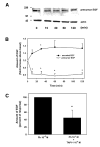
Figure 5. Effect of α1-Adrenergic Agonists on Ectodomain Shedding of EGF
Lacrimal gland pieces were incubated with the α1-adrenergic agonist phenylephrine (10−4 M) for 0–120 minutes. A. Tissue was homogenized and proteins separated on 4–20% polyacrylamide gel and Western blot analysis was performed using an antibody against either EGF or F-actin. B. Western blots from A were scanned, quantified, and the amount of precursor EGF was standardized to the amount of F-actin. After incubation with phenylephrine, the incubation media was collected and the amount of EGF was determined by ELISA using the same antibody used in A (secreted EGF). Data are mean ± SEM of four independent experiments. * indicates statistical significance from 0 time. C. Acini were preincubated with TAPI-1 for 20 minutes prior to stimulation with phenylephrine for 30 minutes. The amount of EGF present in the media was determined by ELISA using the same antibody used in A. Data are mean ± SEM of four independent experiments. * indicates statistical significance from phenylephrine alone.
Figure 6
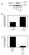
Figure 6. EGF shed by α1-Adrenergic Agonists is Biologically Active and Interacts with the EGF receptor
Lacrimal gland pieces were incubated with either no additions or the α1-adrenergic agonist phenylephrine (10−4 M) for 30 minutes. The conditioned media (c-KRB) was removed and added to acini pretreated with the α1D-adrenergic receptor inhibitor BMY-7378 (10−7 M) for 1 min. The amount of EGF receptor phosphorylation was measured by Western blot analysis. A representative blot is shown in (A). Experiments were quantified and the amount of EGF receptor standardized to actin and shown in (B). Data are mean ± SEM of four independent experiments. Acini were preincubated with AG1478 (10−7 M) for 20 minutes. c-KRB was added for 1 min. Experiments were quantified and the amount of EGF receptor was standardized to actin and shown in (C). Data are mean ± SEM of three independent experiments.
Figure 7
Figure 7. EGF shed by α1-Adrenergic Agonists activates p42/p44 MAPK
Lacrimal gland pieces were incubated with either no additions or the α1-adrenergic agonist phenylephrine (10−4 M) for 30 minutes. The conditioned media (c-KRB) was removed and added to freshly isolated acini pretreated for 20 minutes with BMY-7378 (10−7 M). The amount of p42/p44 MAPK activation was measured after 1 min by Western blot analysis. A representative blot is shown in (A). Experiments were quantified and the amount of phosphorylated p42/p44 MAPK standardized to total p42/p44 MAPK and shown in (B). Data are mean ± SEM of five independent experiments. Acini were preincubated with AG1478 (10−7 M) for 20 minutes before stimulation with c-KRB. Experiments were quantified and the amount of p42/p44 MAPK activation was measured by Western blot analysis and shown in (C). Data are mean ± SEM of three independent experiments.
Figure 8
Figure 8. Biologically Active EGF Shed by by α1-Adrenergic Agonists
Lacrimal gland pieces were incubated with either no additions or the α1-adrenergic agonist phenylephrine (10−4 M) for 30 minutes. The conditioned media (c-KRB) was removed and incubated for 15 min with an EGF neutralizing antibody (20 μg/ml) before addition to freshly isolated acini pretreated for 20 minutes with BMY-7378 (10−7 M). A representative blot is shown in A. Experiments were quantified and the amount of p42/p44 MAPK activation was measured by Western blot analysis and shown in B. Data are mean ± SEM of five independent experiments. As a control for the antibody specificity, exogenous EGF (10−7 M) was preincubated for 15 min with the EGF neutralizing antibody (20 μg/ml) before addition to freshly isolated acini. A representative blot is shown in C. Experiments were quantified and the amount of p42/p44 MAPK activation was measured by Western blot analysis and shown in D. Data are mean ± SEM of five independent experiments.

Comment in

References

    1. Arevalo-Leon LE, Gallardo-Ortiz IA, Urquiza-Marin H, Villalobos-Molina R. Evidence for the role of alpha1D- and alpha1A-adrenoceptors in contraction of the rat mesenteric artery. Vascul Pharmacol. 2003;40:91–96. - PubMed
    1. Arribas J, Coodly L, Vollmer P, Kishimoto TK, Rose-John S, Massague J. Diverse cell surface protein ectodomains are shed by a system sensitive to metalloprotease inhibitors. J Biol Chem. 1996;271:11376–11382. - PubMed
    1. Brown CL, Meise KS, Plowman GD, Coffey RJ, Dempsey PJ. Cell surface ectodomain cleavage of human amphiregulin precursor is sensitive to a metalloprotease inhibitor. Release of a predominant N-glycosylated 43-kDa soluble form. J Biol Chem. 1998;273:17258–17268. - PubMed
    1. Chalothorn D, McCune DF, Edelmann SE, Garcia-Cazarin ML, Tsujimoto G, Piascik MT. Differences in the cellular localization and agonist-mediated internalization properties of the alpha(1)-adrenoceptor subtypes. Mol Pharmacol. 2002;61:1008–1016. - PubMed
    1. Chen J, Acton TB, Basu SK, Montelione GT, Inouye M. Enhancement of the solubility of proteins overexpressed in Escherichia coli by heat shock. J Mol Microbiol Biotechnol. 2002;4:519–524. - PubMed

Publication types

MeSH terms

Substances

LinkOut - more resources