Review
. 2006 Jun;70(2):317-43.
doi: 10.1128/MMBR.00038-05.

Cell wall assembly in Saccharomyces cerevisiae

Affiliations
Review

Cell wall assembly in Saccharomyces cerevisiae

Guillaume Lesage et al. Microbiol Mol Biol Rev. 2006 Jun.

Abstract

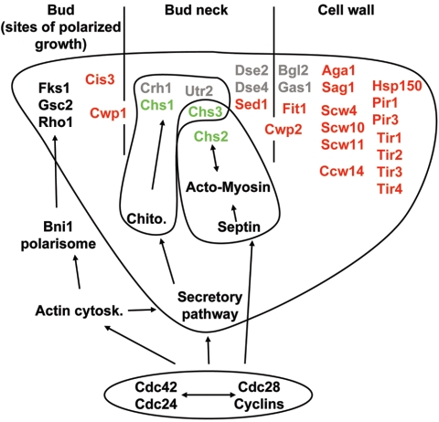
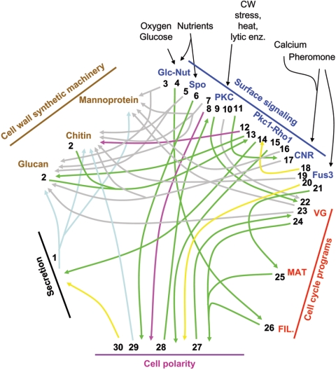
An extracellular matrix composed of a layered meshwork of beta-glucans, chitin, and mannoproteins encapsulates cells of the yeast Saccharomyces cerevisiae. This organelle determines cellular morphology and plays a critical role in maintaining cell integrity during cell growth and division, under stress conditions, upon cell fusion in mating, and in the durable ascospore cell wall. Here we assess recent progress in understanding the molecular biology and biochemistry of cell wall synthesis and its remodeling in S. cerevisiae. We then review the regulatory dynamics of cell wall assembly, an area where functional genomics offers new insights into the integration of cell wall growth and morphogenesis with a polarized secretory system that is under cell cycle and cell type program controls.


Figures

FIG. 1.
FIG. 1.
Schematic representation of cell wall components and their linkages. (A) Architecture of the lateral cell wall. The β-1,3-, β-1,4-, and β-1,6-glucosidic bonds are represented as green, blue, and orange, respectively. Cell wall mannoproteins (CWP) can be linked to the β-1,3-glucan via alkali-sensitive bonds (ASB) or to PIR proteins (PIR) via a disulfide link (SS). GPI cell wall proteins (GPI-CWP) are attached to the β-1,6-glucan through a remnant GPI anchor (GPI Rem.). The links between β-1,3-glucan and β-1,6-glucan or PIR proteins are still uncharacterized (?). Note that the figure qualitatively illustrates the cell wall linkages but does not reflect their stoichiometry. (B) Assembly of cell wall constituents at the septum during bud emergence and at the mother/daughter interface (left and middle panels, respectively) and in the bud scar (right panel). Color representation: lateral cell wall, light gray; plasma membrane, dark gray; chitin ring, pink; chitinous primary septum, yellow; glucan- and mannoprotein-based secondary septum, green; chitinous bud scar, blue. (C) Ascospore wall organization. The glucan-mannoprotein, chitosan, and dityrosine layers are shown in green, blue, and violet, respectively. Chitosan-rich interspore bridges connecting the spores are shown in blue. For clarity the ascus cell wall has been omitted.
FIG. 2.
FIG. 2.
Trafficking and biosynthetic pathways for chitin synthases 2 (A) and 3 (B). The CHS2 and CHS3 genes and their products are represented in red (A) and green (B), respectively. The chitin made by Chs2 is shown in yellow, and that made by Chs3 is shown in pink. The cell wall, the plasma membrane, and septin rings are shown in light and dark gray and as a blue ring, respectively (A and B), and the actin-myosin ring is represented as a hatched gray ring (A). Nucleus, Nuc.; vacuole, Vac.
FIG. 3.
FIG. 3.
Regulation of cell wall gene expression during the cell cycle (A) and in response to external stimuli (B). Boxes contain genes whose expression is controlled by the same pathway or transcription factor. Genes are color coded: black, glucan synthesis; green, chitin synthesis; red, mannoprotein biosynthesis; gray, cell wall remodeling.
FIG. 4.
FIG. 4.
The central role of Rho1 in integrating multiple signals. Rho1 transmits signals from the cell wall sensors and the actin cytoskeleton through GAPs and GEFs to its effectors. Rho1 effectors are colored according to signal input provenance as follows: green, cell wall; red, actin; gray, unknown. Two colors indicate a dual regulation.
FIG. 5.
FIG. 5.
Regulation of transport and localization of proteins involved in cell wall assembly. Proteins are grouped according to their final localization (bud, bud neck, or in the cell wall; note that Cwp1 and Cwp2 show dual localizations) and colored according to their function in cell wall assembly as in Fig. 3. Actin cytosk. actin cytoskeleton; Chito., chitosomes.
FIG. 6.
FIG. 6.
Integrated view of regulatory pathways involved in cell wall biology. Five levels of regulation are represented (cell wall synthetic machinery, brown; surface signaling, blue; cell cycle, red; cell polarity, purple; secretion/endocytosis, black). Interconnections among processes are indicated by arrows, which are numbered from their starting points and colored according to the type of interconnection (regulation of activity, green; expression regulation, gray; spatial regulation, yellow; vesicular transport, blue; undefined regulatory event, pink). Protein transport and glycosylation through the secretory pathway and protein recycling through the endocytic pathway affect the synthesis of glucan, chitin, and mannoprotein (1). The PKC cell integrity pathway is activated by glucan and chitin defects (2). β-1,3-GS and mannoprotein expression is regulated by glucose, nutrients, and oxygen (3). Nutrient availability affects filamentation through Kss1 and cyclin regulation (4). Sporulation regulates GSC2 and CHS3 expression through Ndt80 (5) and affects polarisome function through Kss1 (6). Slt2 acts through Rlm1 on glucan, chitin, and mannoprotein synthesis (7) and on the actin cytoskeleton (8). Pkc1 plays a role in the cell cycle (9). Slt2 acts on the SBF transcription factor (10). Slt2 affects calcineurin signaling through Cch1-Mid1 (11). Pkc1 phosphorylates Chs3 (12). Rho1 is the regulatory subunit of the β-1,3-GS (13). The chitosome is mobilized by the action of Rho1 and Pkc1 on the exocyst and on the secretory pathway, respectively (14). Bni1 is a Rho1 effector (15). The Rho1 effector Skn7 has a role in OCH1 expression (16). GSC2 expression is controlled by the calcineurin-dependent transcription factor Crz1 (17). Pkc1 and Rho1 are recruited at the shmoo tip (18). Pheromone exposure induces CHS1 (19), leads to recruitment of the cell polarity machinery at the shmoo tip (20), and induces a cell cycle arrest (21). The PIR genes are cell cycle regulated (22). Many cell wall genes are expressed in G1 (23). Cdc28 regulates the function of Cdc42 under vegetative growth (24), during mating (25), and during filamentation (26), respectively. The morphogenesis and the cell wall checkpoints affect the cell cycle through the septins and dynactin, respectively (27). Slt2 is regulated by the polarisome (28). Bud neck assembly regulates Chs2 function (29), and cell polarity directs secretion (30). CNR, calcineurin pathway; FIL, filamentous growth program; Fus3, mating pheromone pathway; Glc-Nut, glucose and nutrient sensing; MAT, mating program; PKC, PKC cell integrity pathway; Pkc1-Rho1, signaling through Pkc1 or Rho1 and not through the cell integrity pathway; Spo, sporulation cascade; VG, vegetative growth.

References

    1. Abe, M., H. Qadota, A. Hirata, and Y. Ohya. 2003. Lack of GTP-bound Rho1p in secretory vesicles of Saccharomyces cerevisiae. J. Cell Biol. 162:85-97. - PMC - PubMed
    1. Abeijon, C., and L. Y. Chen. 1998. The role of glucosidase I (Cwh41p) in the biosynthesis of cell wall β-1,6-glucan is indirect. Mol. Biol. Cell 9:2729-2738. - PMC - PubMed
    1. Abramova, N., O. Sertil, S. Mehta, and C. V. Lowry. 2001. Reciprocal regulation of anaerobic and aerobic cell wall mannoprotein gene expression in Saccharomyces cerevisiae. J. Bacteriol. 183:2881-2887. - PMC - PubMed
    1. Adams, D. J. 2004. Fungal cell wall chitinases and glucanases. Microbiology 150:2029-2035. - PubMed
    1. Agarwal, A. K., P. D. Rogers, S. R. Baerson, M. R. Jacob, K. A. T. H. Barker, J. D. Cleary, L. A. Walker, D. G. Nagle, and A. M. Clark. 2003. Genome-wide expression profiling of the response to polyene, pyrimidine, azole, and echinocandin antifungal agents in Saccharomyces cerevisiae. J. Biol. Chem. 278:34998-35015. - PubMed

Publication types

MeSH terms

Substances

LinkOut - more resources