

Immune responses to defined antigens of Mycobacterium bovis in cattle experimentally infected with Mycobacterium kansasii
- PMID: 16760317
- PMCID: PMC1489552
- DOI: 10.1128/CVI.00054-06
Immune responses to defined antigens of Mycobacterium bovis in cattle experimentally infected with Mycobacterium kansasii
Abstract
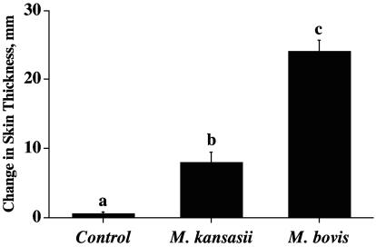
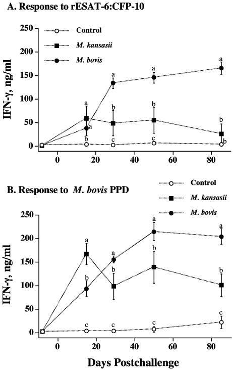
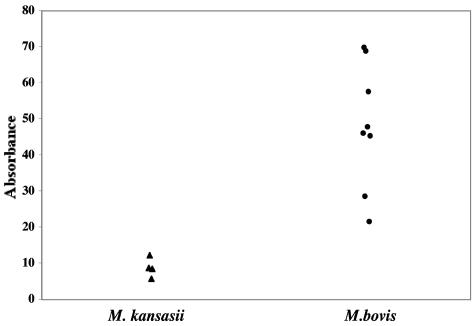
Cross-reactive responses elicited by exposure to nontuberculous mycobacteria often confound the interpretation of antemortem tests for Mycobacterium bovis infection of cattle. The use of specific proteins (e.g., ESAT-6, CFP-10, and MPB83), however, generally enhances the specificity of bovine tuberculosis tests. While genes for these proteins are absent from many nontuberculous mycobacteria, they are present in M. kansasii. Instillation of M. kansasii into the tonsillar crypts of calves elicited delayed-type hypersensitivity and in vitro gamma interferon and nitrite concentration responses of leukocytes to M. avium and M. bovis purified protein derivatives (PPDs). While the responses of M. kansasii-inoculated calves to M. avium and M. bovis PPDs were approximately equivalent, the responses of M. bovis-inoculated calves to M. bovis PPD exceeded their respective responses to M. avium PPD. The gamma interferon and nitrite responses of M. kansasii-inoculated calves to recombinant ESAT-6-CFP-10 (rESAT-6-CFP-10) exceeded corresponding responses of noninoculated calves as early as 15 and 30 days after inoculation, respectively, and persisted throughout the study. The gamma interferon and nitrite responses of M. bovis-inoculated calves to rESAT-6-CFP-10 exceeded the corresponding responses of M. kansasii-inoculated calves beginning 30 days after inoculation. By using a lipoarabinomannan-based enzyme-linked immunosorbent assay, specific serum antibodies were detected as early as 50 days after challenge with M. kansasii. By a multiantigen print immunoassay and immunoblotting, serum antibodies to MPB83, but not ESAT-6 or CFP-10, were detected in M. kansasii-inoculated calves; however, responses to MPB83 were notably weaker than those elicited by M. bovis infection. These findings indicate that M. kansasii infection of calves elicits specific responses that may confound the interpretation of bovine tuberculosis tests.
Figures

References
-
- Alcaide, F., I. Richter, C. Bernasconi, B. Springer, C. Hagenau, R. Schulze-Robbecke, E. Tortoli, R. Martin, E. C. Böttger, and A. Telenti. 1997. Heterogeneity and clonality among isolates of Mycobacterium kansasii: implications for epidemiological and pathogenicity studies. J. Clin. Microbiol. 35:1959-1964. - PMC - PubMed
-
- Arend, S. M., P. de Haas, E. Leyten, I. Rosenkrands, L. Rigouts, P. Andersen, W. Mijs, J. T. van Dissel, and D. van Soolingen. 2005. ESAT-6 and CFP-10 in clinical versus environmental isolates of Mycobacterium kansasii. J. Infect. Dis. 191:1301-1310. - PubMed
-
- Arend, S. M., E. Cerda de Palou, P. de Haas, R. Janssen, M. A. Hoeve, E. M. Verhard, T. H. Ottenhoff, D. van Soolingen, and J. T. van Dissel. 2004. Pneumonia caused by Mycobacterium kansasii in a series of patients without recognised immune defect. Clin. Microbiol. Infect. 10:738-748. - PubMed
-
- Arend, S. M., K. E. van Meijgaarden, K. de Boer, E. C. de Palou, D. van Soolingen, T. H. Ottenhoff, and J. T. van Dissel. 2002. Tuberculin skin testing and in vitro T cell responses to ESAT-6 and culture filtrate protein 10 after infection with Mycobacterium marinum or M. kansasii. J. Infect. Dis. 186:1797-1807. - PubMed
-
- Arend, S. M., T. H. Ottenhoff, P. Andersen, and J. T. van Dissel. 2001. Uncommon presentations of tuberculosis: the potential value of a novel diagnostic assay based on the Mycobacterium tuberculosis-specific antigens ESAT-6 and CFP-10. Int. J. Tuberc. Lung Dis. 5:680-686. - PubMed
Publication types
MeSH terms
Substances
LinkOut - more resources
Full Text Sources
Other Literature Sources
Medical
Miscellaneous
