Comparative Study
. 2006 Jun;13(6):648-54.
doi: 10.1128/CVI.00061-06.

Early antibody responses to experimental Mycobacterium bovis infection of cattle

Affiliations
Comparative Study

Early antibody responses to experimental Mycobacterium bovis infection of cattle

W R Waters et al. Clin Vaccine Immunol. 2006 Jun.

Abstract

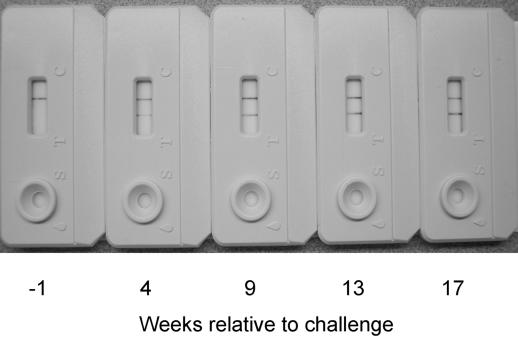
Bovine tuberculosis persists as a costly zoonotic disease in numerous countries despite extensive eradication and control efforts. Sequential serum samples obtained from Mycobacterium bovis-infected cattle were evaluated for seroreactivity to mycobacterial antigens. Animals received M. bovis by aerosol, intratonsil, intranasal, or intratracheal inoculation. Assays included the multiantigen print immunoassay for determination of antigen recognition patterns, immunoblot analysis for sensitive kinetic studies, and the VetTB STAT-PAK test, a novel, rapid test based on lateral-flow technology. Responses to MPB83 were detected for all M. bovis-infected animals regardless of the route or strain of M. bovis used for inoculation. Other less commonly recognized antigens included ESAT-6, CFP-10, and MPB70. Responses to MPB83 were detectable as early as 4 weeks after inoculation, were boosted upon injection of purified protein derivatives for skin testing, and persisted throughout the course of each of the four challenge studies. MPB83-specific immunoglobulin M (IgM) was detected prior to MPB83-specific IgG detection; however, early IgM responses rapidly waned, suggesting a benefit of tests that detect both IgM- and IgG-specific antibodies. The VetTB STAT-PAK test detected responses in sera from 60% (15/25) of the animals by 7 weeks after challenge and detected responses in 96% (24/25) of the animals by 18 weeks. These findings demonstrate the potential for new-generation antibody-based tests for the early detection of M. bovis infection in cattle.


Figures

FIG. 1.
FIG. 1.
Antigen recognition patterns by antibody from cattle inoculated with Mycobacterium bovis via the aerosol route. Cattle received either 105 CFU of M. bovis strain 95-1315 (animals 302, 418a, 425, and 432) or 105 CFU of M. bovis strain HC2005T (animals 293, 300, 408, 410, and 426), and serological reactivity to M. tuberculosis complex antigens was determined by multiantigen immunoprint assay of sequential serum samples. Antigens are indicated in the far-right margin. For each panel of five nitrocellulose strips, animals are indicated in the upper margins, and weeks after challenge are indicated in the lower margins. E6P10 refers to the ESAT-6:CFP10 fusion protein. 16/83 refers to 16-kDa alpha-crystallin/MPB83 fusion protein. PPDb refers to M. bovis PPD. MBCF refers to M. bovis culture filtrate.
FIG. 2.
FIG. 2.
Antigen recognition patterns by antibody from cattle inoculated with Mycobacterium bovis strain 95-1315 via intratonsillar instillation. Serological reactivity to M. tuberculosis complex antigens was determined by multiantigen immunoprint assay of sequential serum samples. Antigens are indicated in the far-right margin. For each panel of five nitrocellulose strips, animals are indicated in the upper margins, and weeks after challenge are indicated in the lower margins. E6P10 refers to the ESAT-6:CFP10 fusion protein. 16/83 refers to the 16-kDa alpha-crystallin/MPB83 fusion protein. PPDb refers to M. bovis PPD. MBCF refers to M. bovis culture filtrate.
FIG. 3.
FIG. 3.
Kinetics of the response to MPB83 by antibodies from cattle inoculated with Mycobacterium bovis via the aerosol route. Cattle received either 105 CFU of M. bovis strain 95-1315 (animals 302, 418a, 425, and 432) or 105 CFU of M. bovis strain HC2005T (animals 293, 300, 408, 410, and 426), and serological reactivity to MPB83 was determined by immunoblot analysis of sequential serum samples. Weeks after challenge are indicated in the upper margin, and animals are indicated in the right margin. In the lower margin, CCT indicates the time point for in vivo administration of purified protein derivatives (i.e., M. avium and M. bovis PPD) for the comparative cervical test (CCT).
FIG. 4.
FIG. 4.
Kinetics of the response to MPB83 by antibodies from cattle inoculated with Mycobacterium bovis strain 95-1315 via intratonsillar instillation. Serological reactivity to MPB83 was determined by immunoblot analysis of sequential serum samples. Weeks after challenge are indicated in the upper margin, and animals are indicated in the right margin. In the lower margin, CFT indicates the time point for the in vivo administration of M. bovis PPD for the caudal fold test (CFT), and CCT indicates the time point for the in vivo administration of purified protein derivatives (i.e., M. avium and M. bovis PPD) for the comparative cervical test (CCT).
FIG. 5.
FIG. 5.
Response kinetics and intensity of reactivity to MPB83 by IgM (A) and IgG (B) in sequential serum samples obtained from cattle inoculated with Mycobacterium bovis via the aerosol route. Semiquantitative densitometry using Scion Image (version beta 4.0.2) was used to determine the intensity of IgM (A) or IgG (B) reactivity to MPB83 printed onto nitrocellulose.
FIG. 6.
FIG. 6.
Vet-TB STAT-PAK test results from sequential serum samples from a calf inoculated with Mycobacterium bovis via aerosol. The upper band of reactivity is an internal positive control indicating mobility of the serum sample and diluent across the membrane. The lower bands of reactivity visualized on samples obtained at 4, 8.5, 13, and 17 weeks after challenge indicate a positive reaction to the test antigens.

References

    1. Bannantine, J., and J. R. Stabel. 2000. HspX is present within Mycobacterium paratuberculosis-infected macrophages and is recognized by sera from some infected cattle. Vet. Microbiol. 76:343-358. - PubMed
    1. Cole, S. T., R. Brosch, J. Parkhill, T. Garnier, C. Churcher, D. Harris, S. V. Gordon, et al. 1998. Deciphering the biology of Mycobacterium tuberculosis from the complete genome sequence. Nature 393:537-544. - PubMed
    1. Corner, L. A., M. A. Stevenson, D. M. Collins, and R. S. Morris. 2003. The re-emergence of Mycobacterium bovis infection in brushtail possums (Trichosurus vulpecula) after localised possum eradication. N. Z. Vet. J. 51(2):73-80. - PubMed
    1. Donnelly, C. A., R. Woodroffe, D. R. Cox, F. J. Bourne, C. L. Cheeseman, R. S. Clifton-Hadley, G. Wei, G. Gettinby, P. Gilks, H. Jenkins, W. T. Johnston, A. M. Le Fevre, J. P. McInerney, and W. I. Morrison. 2005. Positive and negative effects of widespread badger culling on tuberculosis in cattle. Nature 439:843-846. - PubMed
    1. European Economic Community. 1980. EEC directive 80/219, amending directive 64/432 annexe B. Off. J. L047:25-32.

Publication types

MeSH terms

LinkOut - more resources