. 2006 Aug;17(8):3423-34.
doi: 10.1091/mbc.e06-04-0371. Epub 2006 Jun 7.

CP110 cooperates with two calcium-binding proteins to regulate cytokinesis and genome stability

Affiliations

CP110 cooperates with two calcium-binding proteins to regulate cytokinesis and genome stability

William Y Tsang et al. Mol Biol Cell. 2006 Aug.

Abstract

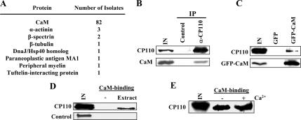
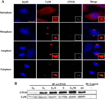
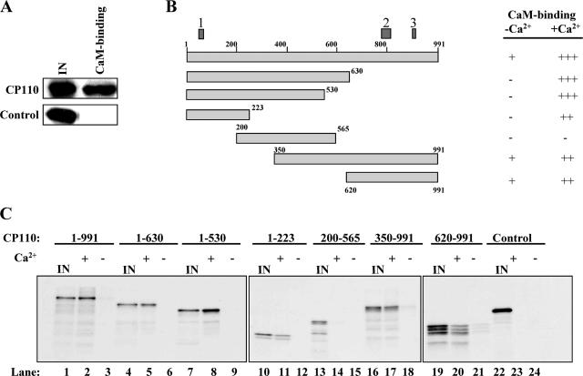
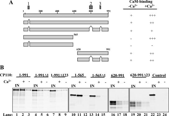
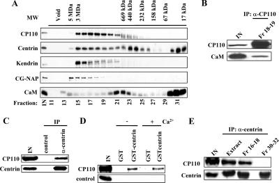
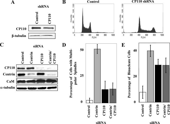
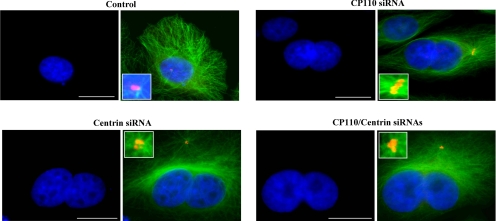
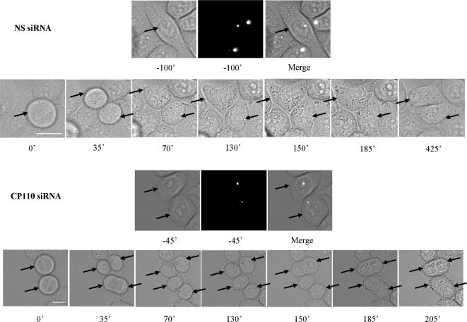
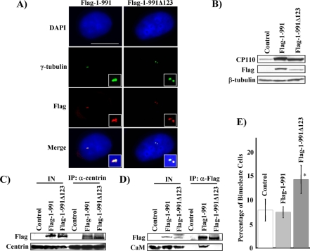
The centrosome is an integral component of the eukaryotic cell cycle machinery, yet very few centrosomal proteins have been fully characterized to date. We have undertaken a series of biochemical and RNA interference (RNAi) studies to elucidate a role for CP110 in the centrosome cycle. Using a combination of yeast two-hybrid screens and biochemical analyses, we report that CP110 interacts with two different Ca2+-binding proteins, calmodulin (CaM) and centrin, in vivo. In vitro binding experiments reveal a direct, robust interaction between CP110 and CaM and the existence of multiple high-affinity CaM-binding domains in CP110. Native CP110 exists in large (approximately 300 kDa to 3 MDa) complexes that contain both centrin and CaM. We investigated a role for CP110 in CaM-mediated events using RNAi and show that its depletion leads to a failure at a late stage of cytokinesis and the formation of binucleate cells, mirroring the defects resulting from ablation of either CaM or centrin function. Importantly, expression of a CP110 mutant unable to bind CaM also promotes cytokinesis failure and binucleate cell formation. Taken together, our data demonstrate a functional role for CaM binding to CP110 and suggest that CP110 cooperates with CaM and centrin to regulate progression through cytokinesis.


Figures

Figure 1.
Figure 1.
CaM interacts with CP110 in vivo. (A) The number of instances that each annotated protein was identified in the yeast two-hybrid screen is shown. (B) Western blotting of endogenous CP110 and CaM after immunoprecipitation with anti-CP110 antibody or control (anti-calnexin) antibody from 293T cell extracts. IN, input. (C) Western blotting of endogenous CP110 and recombinant GFP-CaM after immunoprecipitation with anti-GFP antibody using extracts from GFP-CaM- (GFP-CaM) and GFP-expressing cells (GFP). (D) Western blot detection of endogenous CP110 and control (giantin) after binding of CaM agarose with 293 extracts (extract) or with lysis buffer (−). (E) Western blot detection of endogenous CP110 after binding of CaM agarose with 293 extracts supplemented with either EGTA (−Ca2+) or calcium (+Ca2+).
Figure 2.
Figure 2.
Colocalization and coimmunoprecipitation of CP110 and CaM across the cell cycle. (A) U2OS cells were processed for immunofluorescence with anti-CaM antibody (red) and anti-CP110 antibody (green). DNA was stained with DAPI (blue). A representative interphase, metaphase, anaphase, and telophase cell are illustrated. Yellow color in merged images indicates substantial colocalization of CP110 and CaM. Insets show magnified view of centrosomes. Bar, 10 μm; insets, 3 μm. (B) T98G cells were synchronized by serum starvation and restimulated with serum to initiate cell cycle reentry. Cell lysates from different cell cycle stages were collected. Western blots of endogenous CP110 and CaM after immunoprecipitation with anti-CP110 antibody or control (anti-calnexin) antibody.
Figure 3.
Figure 3.
CP110 interacts with CaM through multiple domains in vitro. (A) Binding of in vitro–translated 35S-labeled CP110 or 35S-labeled control (luciferase) with CaM agarose beads. The binding was carried out in a lysis buffer without the addition of EGTA or calcium. (B) Schematic representation of CP110 truncation mutants used to map CaM-binding domains. The three boxes (1, 2, and 3) denote the three CaM-binding domains predicted by the Calmodulin Target Database. The strength of CaM-binding was quantitated with a densitometer and is categorized as strong (+++), intermediate (++), weak (+), or none (−). (C) In vitro binding assays. The results were summarized in B.
Figure 4.
Figure 4.
CP110 CaM-binding mutants 1-565Δ1 and 1-991Δ123 interact poorly with CaM in vitro. (A) Schematic representation of CP110 truncation mutants and CaM-binding domains. The three boxes (1, 2, and 3) denote the three CaM-binding domains predicted by the Calmodulin Target Database. The strength of CaM-binding was quantitated with a densitometer and is categorized as strong (+++), intermediate (++), weak (+), or none (−). (B) In vitro binding assays. The results were summarized in A.
Figure 5.
Figure 5.
Centrin interacts with CP110 in vivo and cofractionates with CP110 and CaM in high-molecular-weight complexes. (A) Cell extract was chromatographed on a Superose 6 gel filtration column, and the resulting fractions were Western blotted with antibodies against CP110, centrin, kendrin, CG-NAP, or CaM. Estimated molecular weights are indicated at the top of the panel. (B) Western blotting of endogenous CP110 and CaM after immunoprecipitation with anti-CP110 antibody using fractions 18–19 (Fr 18–19) from the Superose 6 column. (C) Western blot of endogenous CP110 and centrin after immunoprecipitation with anti-centrin antibody or control (anti-calnexin) antibody using extracts from 293T cells. (D) Western blotting of endogenous CP110 and control (giantin) after binding of either GST or GST-centrin prebound to glutathione agarose beads with 293T extracts. The buffer used for prebinding and the 293T extracts were supplemented with either EGTA (−Ca2+) or calcium (+Ca2+). (E) Western blotting of endogenous CP110 and centrin after immunoprecipitation with anti-centrin antibody using 293T cell extract (Extract), fractions 16–18 (Fr 16–18), or fractions 30–32 (Fr 30–32) from the Superose 6 column.
Figure 6.
Figure 6.
RNAi-mediated suppression of CP110 induces polyploidy and results in the formation of binucleate cells. (A) Western blot detection of CP110 proteins in U2OS cells transfected with a control or CP110 shRNA expression vector. β-tubulin was used as a loading control. (B) FACS analysis of control or CP110 shRNA-expressing cells. (C) Western blotting of CP110, centrin, and CaM in HeLa cells treated with control, centrin, CP110, or centrin and CP110 siRNAs. α-tubulin was used as a loading control. (D) The percentages of cells with multipolar spindles were determined. About 50 mitotic cells were scored for each condition, and the experiments were repeated at least twice. (E) The percentages of binucleate cells were determined. About 200 cells were scored for each condition, and the experiments were repeated at least three times.
Figure 7.
Figure 7.
Ablation of CP110 and centrin results in the formation of binucleate cells. HeLa cells transiently transfected with control, CP110, centrin, or CP110 and centrin siRNAs stained with antibodies to α-tubulin (green), γ-tubulin (red), and with DAPI (blue). Only DAPI and merged images are shown. Bar, 10 μm; insets, 2 μm.
Figure 8.
Figure 8.
Cells depleted of CP110 exhibit a late cytokinesis defect. HeLa cells transfected with rhodamine-labeled and nonspecific control siRNA oligonucleotides or rhodamine-labeled and CP110 siRNA oligonucleotides were observed by DIC time-lapse videomicroscopy. At least 80 mitotic events were scored for each condition. A representative cell treated with control and two representative cells treated with CP110 siRNAs are illustrated. Times from the onset of metaphase are indicated. The fluorescent images were taken before mitosis. Arrows indicate transfected cells and their daughter cells. Bar, 10 μm.
Figure 9.
Figure 9.
Ectopic expression of a CP110 CaM-binding mutant in vivo results in binucleate cell formation. (A) U2OS cells transiently transfected with a Flag-1-991 or Flag-1-991Δ123 expression vector stained with antibodies to γ-tubulin (green), Flag (red), and with DAPI (blue). Bar, 10 μm; insets: 2 μm. (B) Western blot detection of CP110 proteins in HeLa cells transfected with a control, Flag-1-991 or Flag-1-991Δ123 expression vector. The blots labeled CP110 and Flag were probed with anti-CP110 and anti-Flag antibodies, respectively. β-tubulin was used as a loading control. (C) Western blotting of Flag-CP110 and endogenous centrin after immunoprecipitation with anti-centrin antibody using 293T cell extract expressing Flag (control), Flag-1-991 or Flag-1-991Δ123 proteins. IN, input. (D) Western blotting of Flag-CP110 and endogenous CaM after immunoprecipitation with anti-Flag antibody using 293T cell extract expressing Flag (control), Flag-1-991 or Flag-1-991Δ123 proteins. IN, input. (E) HeLa cells were transfected with a G418-resistant marker along with a control, Flag-1-991 or Flag-1-991Δ123 expression vector. The percentages of binucleate cells were determined after 5–6 d of selection in the presence of G418. About 500 cells were scored for each condition, and the experiments were repeated at least twice. Asterisk denotes that the percentage of binucleate cells resulting from expression of the mutant (Flag-1-991Δ123) is significantly higher than that of control (p < 0.01) or wild-type (p < 0.01) based on a two-tailed Student's t test.

Similar articles

Cited by

References

    1. Andersen J. S., Wilkinson C. J., Mayor T., Mortensen P., Nigg E. A., Mann M. Proteomic characterization of the human centrosome by protein correlation profiling. Nature. 2003;426:570–574. - PubMed
    1. Badano J. L., Teslovich T. M., Katsanis N. The centrosome in human genetic disease. Nat. Rev. Genet. 2005;6:194–205. - PubMed
    1. Chen Z., Indjeian V. B., McManus M., Wang L., Dynlacht B. D. CP110, a cell cycle-dependent CDK substrate, regulates centrosome duplication in human cells. Dev. Cell. 2002;3:339–350. - PubMed
    1. Chin D., Means A. R. Calmodulin: a prototypical calcium sensor. Trends Cell Biol. 2000;10:322–328. - PubMed
    1. Debec A. Haploid cell cultures of Drosophila melanogaster. Nature. 1978;274:255–256. - PubMed

Publication types

MeSH terms