. 2006 Aug;17(8):3543-56.
doi: 10.1091/mbc.e06-04-0322. Epub 2006 Jun 7.

Stable CpG hypomethylation of adipogenic promoters in freshly isolated, cultured, and differentiated mesenchymal stem cells from adipose tissue

Affiliations

Stable CpG hypomethylation of adipogenic promoters in freshly isolated, cultured, and differentiated mesenchymal stem cells from adipose tissue

Agate Noer et al. Mol Biol Cell. 2006 Aug.

Abstract

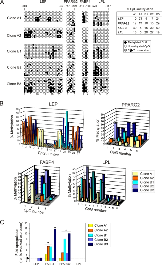
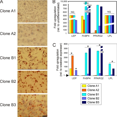
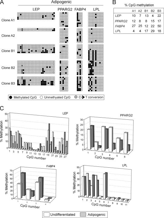
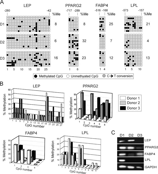
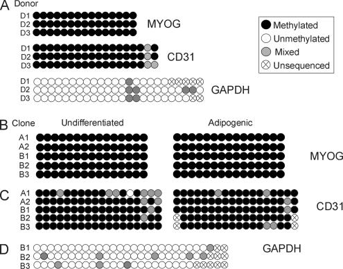
Mesenchymal stem cells from adipose tissue can differentiate into mesodermal lineages. Differentiation potential, however, varies between clones of adipose stem cells (ASCs), raising the hypothesis that epigenetic differences account for this variability. We report here a bisulfite sequencing analysis of CpG methylation of adipogenic (leptin [LEP], peroxisome proliferator-activated receptor gamma 2 [PPARG2], fatty acid-binding protein 4 [FABP4], and lipoprotein lipase [LPL]) promoters and of nonadipogenic (myogenin [MYOG], CD31, and GAPDH) loci in freshly isolated human ASCs and in cultured ASCs, in relation to gene expression and differentiation potential. Uncultured ASCs display hypomethylated adipogenic promoters, in contrast to myogenic and endothelial loci, which are methylated. Adipogenic promoters exhibit mosaic CpG methylation, on the basis of heterogeneous methylation between cells and of variation in the extent of methylation of a given CpG between donors, and both between and within clonal cell lines. DNA methylation reflects neither transcriptional status nor potential for gene expression upon differentiation. ASC culture preserves hypomethylation of adipogenic promoters; however, between- and within-clone mosaic methylation is detected. Adipogenic differentiation also maintains the overall CpG hypomethylation of LEP, PPARG2, FABP4, and LPL despite demethylation of specific CpGs and transcriptional induction. Furthermore, enhanced methylation at adipogenic loci in primary differentiated cells unrelated to adipogenesis argues for ASC specificity of the hypomethylated state of these loci. Therefore, mosaic hypomethylation of adipogenic promoters may constitute a molecular signature of ASCs, and DNA methylation does not seem to be a determinant of differentiation potential of these cells.


Figures

Figure 1.
Figure 1.
CpG-specific methylation level at the LEP, PPARG2, FABP4, and LPL promoters in cultured ASCs. (A) Distribution of CpGs in each promoter region examined. Numbers indicate nucleotide number upstream of the ATG translational start site (see Supplemental Figure S1 for sequences). Tick marks indicate the position of each CpG. (B–E) Percentage of 5′–3′ CpG methylation determined by bisulfite sequencing at indicated promoters in polyclonal populations of cultured undifferentiated ASCs. Number 1 refers to the 5′-most CpG.
Figure 2.
Figure 2.
Adipogenic loci are hypomethylated in cultured ASC clones, irrespective of gene expression. (A) Bisulfite analysis of LEP, PPARG2, FABP4, and LPL in five ASC clones from two donors (A and B clones, respectively). Table shows the percentage of global CpG methylation (● in A) at each locus for each clone. (B) Proportion of individual methylated CpGs at the LEP, PPARG2, FABP4, and LPL promoter in each clone. (C) QRT-PCR analysis of expression of LEP, PPARG2, FABP4, and LPL in undifferentiated ASC clones, relative to the lowest expressing clone (level 1) for a given gene. Asterisk (*) indicates a statistical difference in expression at the p < 0.01 level (t test) relative to the weakest expressing clone (level 1).
Figure 3.
Figure 3.
Adipogenic differentiation of ASC clones. (A) Morphological evidence of differentiation after 3 wk of adipogenic stimulation (Oil Red-O staining). Bar, 50 μm. (B) QRT-PCR analysis of expression of indicated genes in each ASC clone after 3 wk of differentiation, relative to expression level in the same but undifferentiated clone. (C) Gene expression analysis as in B, but expressed relative to the lowest expressing clone for a given gene. All samples were analyzed in triplicates. NQ indicates gene expression, but level was not quantified due the absence of expression in undifferentiated ASCs (value was arbitrarily set on graph). *p < 0.005 (t test) for all transcripts with an expression level >10-fold relative to the weakest expressing clone (level 1), and p < 0.001 for genes indicated in the text.
Figure 4.
Figure 4.
Bisulfite sequencing analysis of DNA methylation of LEP, PPARG2, FABP4, and LPL promoters in ASC clones after adipogenic differentiation. (A) Bisulfite analysis. (B) Percentage of global CpG methylation (◇ in A) at each promoter for each clone. (C) Average percentage of individual CpG methylation, across all clones, in undifferentiated ASCs and after adipogenic differentiation. Statistical analysis (paired t tests) of differences in percentage methylation is provided in the text.
Figure 5.
Figure 5.
DNA methylation analysis of the LEP promoter in differentiated human SGBS adipocytes. (A) Bisulfite sequencing analysis. (B) Percentage of individual CpG methylation in adipogenic-differentiated ASCs (pool of all clones shown in A) and in SGBS adipocytes. (C) Endpoint RT-PCR analysis of LEP and GAPDH expression in differentiated SGBS adipocytes.
Figure 6.
Figure 6.
DNA methylation analysis of LEP, PPARG2, FABP4, and LPL in freshly isolated, uncultured ASCs. (A) Bisulfite analysis of CpG methylation in ASCs from three donors (D1–D3). Percentage of overall CpG methylation (%Me; ●) is shown. (B) Proportions of individual methylated CpGs at each promoter and or each donor. CpG numbers are indicated, no. 1 being the 5′ most CpG. Note that analysis of 10 bacterial clones for each donor was barely sufficient for statistical comparisons. (C) Endpoint RT-PCR analysis of expression of indicated genes in ASCs purified from donors D1, D2, and D3.
Figure 7.
Figure 7.
Comparison of CpG methylation profiles in uncultured versus cultured ASCs. Average percentages of methylation of individual CpGs in the LEP, PPARG2, FABP4, and LPL promoters in freshly isolated ASCs from all three donors (uncultured) and across all five undifferentiated ASC clones (cultured) are shown.
Figure 8.
Figure 8.
DNA methylation analysis of MYOG, CD31, and GAPDH in uncultured and cultured ASCs. (A) Analysis of ASCs purified from donors D1–D3 resulting from direct sequencing of PCR products after bisulfite conversion. Representative sequences are shown in Supplemental Figure S3. (B and C) Analysis of MYOG (B) and CD31 (C) methylation in undifferentiated and adipogenic-differentiated ASC clones. (D) Analysis of GAPDH methylation in undifferentiated ASC clones B1–B3. The “mixed” methylation pattern is due to the analysis of mixed cell populations because PCR products resulting from bisulfite conversion were not cloned.
Figure 9.
Figure 9.
Bisulfite sequencing analysis of CpG methylation of LEP in primary human nonadipocytic cells. Analysis of uncultured PBLs and T-cells. Percentage of global methylation (●) is shown. CpG numbers are shown, with no. 1 being the 5′-most CpG.

References

    1. Antequera F. Structure, function and evolution of CpG island promoters. Cell. Mol. Life Sci. 2003;60:1647–1658. - PMC - PubMed
    1. Ayyanathan K., Lechner M. S., Bell P., Maul G. G., Schultz D. C., Yamada Y., Tanaka K., Torigoe K., Rauscher F. J., III, et al. Regulated recruitment of HP1 to a euchromatic gene induces mitotically heritable, epigenetic gene silencing: a mammalian cell culture model of gene variegation. Genes Dev. 2003;17:1855–1869. - PMC - PubMed
    1. Azuara V. Chromatin signatures of pluripotent cell lines. Nat. Cell Biol. 2006;8:532–538. - PubMed
    1. Ballas N., Grunseich C., Lu D. D., Speh J. C., Mandel G. REST and its corepressors mediate plasticity of neuronal gene chromatin throughout neurogenesis. Cell. 2005;121:645–657. - PubMed
    1. Bey L., Etienne J., Tse C., Brault D., Noe L., Raisonnier A., Arnault F., Hamilton M. T., Galibert F. Cloning, sequencing and structural analysis of 976 base pairs of the promoter sequence for the rat lipoprotein lipase gene. Comparison with the mouse and human sequences. Gene. 1998;209:31–38. - PubMed

Publication types