. 2006 Aug;17(8):3446-55.
doi: 10.1091/mbc.e06-02-0102. Epub 2006 Jun 7.

A role for the mitogen-activated protein kinase kinase kinase 1 in epithelial wound healing

Affiliations

A role for the mitogen-activated protein kinase kinase kinase 1 in epithelial wound healing

Maoxian Deng et al. Mol Biol Cell. 2006 Aug.

Abstract

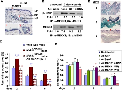
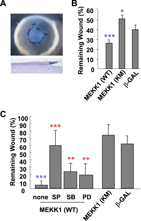
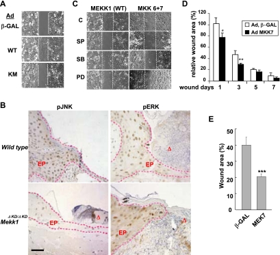
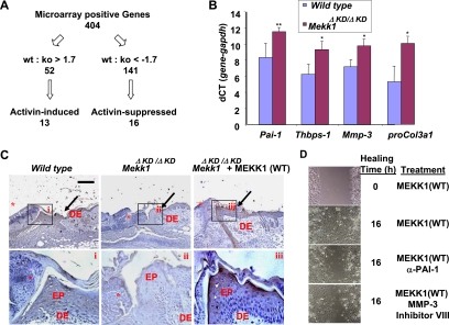
The mitogen-activated protein kinase kinase (MEK) kinase 1 (MEKK1) mediates activin B signals required for eyelid epithelium morphogenesis during mouse fetal development. The present study investigates the role of MEKK1 in epithelial wound healing, another activin-regulated biological process. In a skin wound model, injury markedly stimulates MEKK1 expression and activity, which are in turn required for the expression of genes involved in extracellular matrix (ECM) homeostasis. MEKK1 ablation or down-regulation by interfering RNA significantly delays skin wound closure and impairs activation of Jun NH2-terminal kinases, induction of plasminogen activator inhibitor (PAI)-1, and restoration of cell-cell junctions of the wounded epidermis. Conversely, expression of wild-type MEKK1 accelerates reepithelialization of full-thickness skin and corneal debridement wounds by mechanisms involving epithelial cell migration, a cell function that is partially abolished by neutralizing antibodies for PAI-1 and metalloproteinase III. Our data suggest that MEKK1 transmits wound signals, leading to the transcriptional activation of genes involved in ECM homeostasis, epithelial cell migration, and wound reepithelialization.


Figures

Figure 1.
Figure 1.
MEKK1 is required for optimal skin wound closure. (A) Skin from Mekk1+/ΔKD mice was stained with X-Gal. Tissue sections were examined for blue X-Gal–positive cells accumulating in the epidermis (i) and hair follicles (ii), as indicated. DE, dermis; EP, epithelium; HF, hair follicles. Bar, 50 μm. (B) Wounds on wild-type mice were either uninfected or infected by adenoviruses for GFP and MEKK1 siRNA. Protein lysates from normal and day 3-wounded skin tissues were subjected to immunoprecipitation using α-MEKK1, followed by an in vitro kinase assay using GST-JNKK1 as a substrate (top) and Western blotting using α-MEKK1 (bottom). The -fold induction of MEKK1 expression and kinase activity were relative to those in unwounded skin tissues. (C) Full-thickness excision wounds were created on the dorsal side of wild-type and Mekk1ΔKDKD mice. The Mekk1ΔKDKD wounds were either uninfected or infected with Ad MEKK1(WT). (D) Wounds on wild-type mice were either uninfected or subjected to infection by adenoviruses for β-Gal, MEKK1(WT), MEKK1(KM), GFP and MEKK1 siRNA. Wound areas were measured after various days of injury and are presented relative to the original wound size. The results represent the mean ± SD of at least 16 wounds of each condition. The means of the remaining wound area of each group were analyzed by the Student's t test. ∗∗∗p < 0.001, ∗∗p < 0.01, and ∗p < 0.05 are considered statistically significant. Comparisons of wounds between wild-type versus Mekk1ΔKDKD are labeled in blue; uninfected versus Ad-infected mice of the same genotype are labeled in red. (E) Adenoviruses for β-Gal were injected to the full-thickness skin wounds of wild-type mice. The wound tissues were isolated at various days after injury and subjected to X-Gal staining. Sections of the stained tissues were counterstained by hematoxylin and photographed and the blue β-Gal–positive cells could be identified in the wound tissues. Arrows point at the epidermis.
Figure 2.
Figure 2.
The MEKK1–JNK pathway stimulates corneal reepithelialization and wound healing. (A) β-Gal adenoviruses were applied as eyedrops to the corneal debridement wounds in mice. At 24 h after injury, the eye tissues were subjected to whole mount X-Gal staining (top). The stained tissues were sectioned and counterstained by hematoxylin (bottom) and the blue β-Gal–positive cells could be easily identified in the wounding edge epithelium. (B) Adenoviruses for β-Gal, MEKK1(WT), and MEKK1(KM) were used to infect corneal debridement wounds created in vivo on mouse eyes. The wound areas were measured at 24 h after injury and were calculated relative to the original wounds. Remaining wound areas were compared with those in Ad β-Gal–infected samples. (C) Corneal debridement wounds created on eyes maintained in ex vivo organ culture system were exposed to adenoviruses as indicated, in the presence or absence of MAPK inhibitors SP600125 (SP), SB202190 (SB), and PD98059 (PD) in the culture media. The wound areas were measured 36 h post injury and were calculated relative to the original wounds, or, in the case when inhibitors were used, relative to the wounds the absence of inhibitors. All data represent mean ± SD of at least eight wounds, and statistical analyses were carried out by Student's t test. The p values of ∗∗∗p < 0.001, ∗∗p < 0.01, and ∗p < 0.05 are considered significantly different from the wounds of Ad β-Gal–infected eyes (in blue) or from the same Ad-infected wounds in the absence of the inhibitors (in red).
Figure 3.
Figure 3.
Activation of MEKK1 and JNK is sufficient for keratinocyte migration and skin wound closure. (A) Epidermal keratinocytes were infected by adenoviruses as indicated. The cells were subjected to in vitro wound healing assays and were photographed at 24 h after injury. (B) Skin wound tissues at day 3 after injury were subjected to immunohistochemistry staining for phosphorylated JNK (pJNK) and ERK (pERK), and the positive (brown color) staining was evident in the epidermis of wild-type mice. The epithelium in the wounds is marked by red dotted lines, and pJNK-positive cells were markedly reduced in the epithelium of Mekk1ΔKDKD mice. EP, epithelium and the triangle (Δ) indicates wound exudates. Bar, 50 μm. (C) Cells infected with Ad MEKK1(WT), and Ad MKK6 and Ad MKK7, as indicated, were subjected to in vitro wound healing assays in the absence and presence of various MAPK inhibitors and photographed at 24 h after injury. (D) Full-thickness skin wounds were generated on the dorsal side of wild-type mice. The wounds were exposed to adenoviruses for β-Gal and MKK7. Wound areas were measured at various days after injury, as indicated. (E) Corneal debridement wounds created on eyes maintained in ex vivo organ culture system were exposed to adenoviruses. The wound areas were measured 36 h post injury. The results represent mean ± SD of at least 12 wounds and were analyzed by the Student's t test, relative to Ad β-Gal–infected tissues. ∗∗∗p < 0.001, ∗∗p < 0.01, and ∗p < 0.05 are considered significant. C, no inhibitor; PD, PD98059, an inhibitor of ERK; SB, SB202190, an inhibitor of p38; and SP, SP600125, an inhibitor of JNK.
Figure 4.
Figure 4.
The induction of PAI-1 in the wound epidermis is dependent on MEKK1. (A) Summary of the microarray data. Of the MEKK1-dependent genes, 13 were induced and 16 suppressed by activin B treatment. Cultured epidermal keratinocytes were deprived of growth factors for 24 h, followed by exposure to 5 ng/ml activin B for 12 h or at the indicated times. (B) RNA isolated from epidermal keratinocytes of wild-type and Mekk1ΔKDKD mice were analyzed by real-time PCR for Pai-1, Thbps-1, Mmp-3, Procol3a1, and Gapdh. (C) Full-thickness skin wounds at days 1, 3, 5, and 7 were subjected to immunohistochemistry using anti-PAI-1. Increased PAI-1, characterized as diffused staining in brown color, was observed in the epidermis of the wounds of wild-type mice (i) or in Mekk1ΔKDKD infected by Ad MEKK1(WT) (iii), but it was almost undetectable in that of the Mekk1ΔKDKD (ii). Pictures were taken on skin tissues at day 5 of wounding, although similar PAI-1 expression pattern was observed in tissues at other wounding days. Bar, 250 μm. DE, dermis; EP, epidermis; ∗, exudates; and arrows, wounding edge. (D) In vitro wound healing assay of Ad MEKK1(WT)-infected HaCAT human keratinocytes with or without treatment with 5 μg/ml anti-PAI-1 and 10 μM MMP-3 inhibitor VIII before the wounding. Pictures were taken at 0 and 16 h.
Figure 5.
Figure 5.
Lack of cell–cell junction formation in the wound epidermis of Mekk1ΔKDKD mice at late stage of healing. Morphological examination of the wounding edge epidermis at various days after injury. The wound tissues were examined under either phase-contrast microscopy (A) after H&E staining, and pictures were taken at 1000× magnification (bar, 10 μm), or electron microscopy (B), which was done on day 6 wounds. At early stage (day 3), both wild-type and Mekk1ΔKDKD mice show an apparent increase in intercellular spaces in the wounding edge epithelium. At late stage (days 6 and 10), only the Mekk1ΔKDKD mice display the spaces, whereas wild-type mice show tight cell–cell contact and the formation of desmosomes, similar to those present in normal unwounded skin. CP, cytoplasm; NE, nucleus. Bar, 10 μm.

Similar articles

Cited by

References

    1. Albo D., Berger D. H., Vogel J., Tuszynski G. P. Thrombospondin-1 and transforming growth factor beta-1 upregulate plasminogen activator inhibitor type 1 in pancreatic cancer. J. Gastrointest. Surg. 1999;3:411–417. - PubMed
    1. Ashcroft G. S., et al. Mice lacking Smad3 show accelerated wound healing and an impaired local inflammatory response. Nat. Cell Biol. 1999;1:260–266. - PubMed
    1. Bamberger C., Scharer A., Antsiferova M., Tychsen B., Pankow S., Muller M., Rulicke T., Paus R., Werner S. Activin controls skin morphogenesis and wound repair predominantly via stromal cells and in a concentration-dependent manner via keratinocytes. Am. J. Pathol. 2005;167:733–747. - PMC - PubMed
    1. Bosch M., Serras F., Martin-Blanco E., Baguna J. JNK signaling pathway required for wound healing in regenerating Drosophila wing imaginal discs. Dev. Biol. 2005;280:73–86. - PubMed
    1. Chan J. C., Duszczyszyn D. A., Castellino F. J., Ploplis V. A. Accelerated skin wound healing in plasminogen activator inhibitor-1-deficient mice. Am. J. Pathol. 2001;159:1681–1688. - PMC - PubMed

Publication types

MeSH terms

LinkOut - more resources