Comparative Study
. 2006 Jun 7;26(23):6288-97.
doi: 10.1523/JNEUROSCI.0768-06.2006.

Possible origins of the complex topographic organization of motor cortex: reduction of a multidimensional space onto a two-dimensional array

Affiliations
Comparative Study

Possible origins of the complex topographic organization of motor cortex: reduction of a multidimensional space onto a two-dimensional array

Tyson N Aflalo et al. J Neurosci. .

Abstract

We propose that some of the features of the topographic organization in motor cortex emerge from a competition among several conflicting mapping requisites. These competing requisites include a somatotopic map of the body, a map of hand location in space, and a partitioning of cortex into regions that emphasize different complex, ethologically relevant movements. No one type of map fully explains the topography; instead, all three influences (and perhaps others untested here) interact to form the topography. A standard algorithm (Kohonen network) was used to generate an artificial motor cortex array that optimized local continuity for these conflicting mapping requisites. The resultant hybrid map contained many features seen in actual motor cortex, including the following: a rough, overlapping somatotopy; a posterior strip in which simpler movements were represented and more somatotopic segregation was observed, and an anterior strip in which more complex, multisegmental movements were represented and the somatotopy was less segregated; a clustering of different complex, multisegmental movements into specific subregions of cortex that resembled the arrangement of subregions found in the monkey; three hand representations arranged on the cortex in a manner similar to the primary motor, dorsal premotor, and ventral premotor hand areas in the monkey; and maps of hand location that approximately matched the maps observed in the monkey.


Figures

Figure 1.
Figure 1.
Eight different possible organizations to the lateral motor cortex in the monkey brain. A, A sequence of body parts arranged vertically with the feet in a medial location and the mouth in a lateral location (Fritsch and Hitzig, 1870; Foerster, 1936; Fulton, 1938). B, A subsequent refinement of the map including a horizontal component to the organization, in which the axial musculature is represented anteriorly and the distal musculature posteriorly (Woolsey et al., 1952). C, Another proposed organization in which a posterior hand representation is partly surrounded by an arm representation (Kwan et al., 1978; Park et al., 2001). D, Division of the precentral gyrus into two strips: primary motor cortex that tended to represent individual body parts and the more complex premotor cortex (Campbell, 1905). E, Partitioning of premotor cortex into a dorsal and ventral sector, each sector subdivided into a caudal and a rostral part (Matelli et al., 1985; Preuss et al., 1996). F, Three regions in the lateral motor strip that project directly to the hand portion of the spinal cord: the primary motor hand area, the ventral premotor hand area, and the dorsal premotor hand area (Dum and Strick, 2002, 2005). G, Rough map of the height of the hand as evoked by electrical stimulation. Data from one monkey (Graziano et al., 2005). Upper hand locations are in a ventral location, and lower hand locations are in a dorsal location. Within the arm–leg region of the somatotopy, the mapping of the hand height breaks down. H, Data from the same monkey as in G but now showing the lateral position of hand evoked by electrical stimulation. I, Cortical patches that, when stimulated, result in different ethologically relevant movements. The details differ among monkeys, but the overall pattern is consistent. Data from one monkey (Graziano et al., 2005). A.S., Arcuate sulcus; C.S., central sulcus; F4/F5, fields 4 and 5 of motor cortex; M1, primary motor cortex; PMd, dorsal promotor area; PMv, ventral premotor area; PMDc, dorsal premotor cortex, caudal division; PMDr, dorsal premotor cortex, rostral division; SMA, supplementary motor area.
Figure 2.
Figure 2.
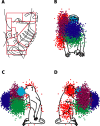
Some movement definitions used in the motor cortex model. A, Woolsey’s map of the somatotopic organization in monkey motor cortex (Woolsey et al., 1952), overlaid with blocked regions showing the schematized somatotopy used in the present motor cortex model. B–D, Three views of a schematized monkey showing the hand locations defined for different ethological categories of movement. Light blue, Hand-to-mouth; dark blue, reach; red, defense; green, central space/manipulation; pink with black border, climbing.
Figure 3.
Figure 3.
Final state of the self-organizing map model. In this learning set, the different movement types were presented to the network in equal proportion. A, The initial somatotopic (Init. Somat.) arrangement of body parts before reorganization. B–K, Somatotopic arrangement of the 10 body parts after reorganization. L, Arrangement of the five ethological categories after reorganization. In L only, light blue, hand-to-mouth; dark blue, reach; red, defense; green, central space/manipulation; pink, climbing. Regions of overlap have intermediate colors. M–O, Maps of hand location after reorganization. X, Hand height, with warm colors indicating greater height; Y, lateral location of hand, with warm colors indicating more lateral locations; Z, distance of hand from body along line of sight, with warm colors indicating more distant locations.
Figure 4.
Figure 4.
Alternate version of the self-organizing map model. In this learning set, different movement types were presented to the network in unequal proportions. The central-space/manipulation movements were increased in proportion by 50%, the reaching movements were decreased by 20%, the hand-in-lower space movements were increased by 50%, and the chewing movements and the leg and foot movements were decreased by 20%. A similar result was obtained here as in Figure 3.
Figure 5.
Figure 5.
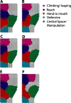
Six variants of the self-organizing map model. Despite the variations in initial state, the results were similar. In each case, the model was the same as shown in Figure 3, except as follows. A, Mean hand location for each of the five movement types was shifted in a random direction by 5 cm. B, The SD of hand location for each of the five movement types was doubled. C–F, The proportions of the movement types in the input array were altered randomly by 20%. In one variant (F), the climbing zone moved to a more posterior region and the reaching zone moved to the dorsal anterior corner of the array. This variant was therefore similar but not identical to the others. This result shows that changing the proportions of the movement types can cause small variations in the final results, but the essential topography remains similar.
Figure 6.
Figure 6.
Usage-dependent changes in the map. The model was trained on the same movement set used in Figure 3 and then trained again on a second movement set. A, Final state of map model when the second movement set was the same as the first. The representation of one ethological movement category, central-space/manipulation, is shown. B, Final state of the map model when the second movement set was the same as the first except that the number of central-space/manipulation movements was increased by 50%. C, Final state of the map model when the second movement set was the same as the first except that the number of central-space/manipulation movements was decreased by 50%.
Figure 7.
Figure 7.
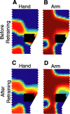
Effect of a “lesion” on the map model. A, Final state of the hand representation using parameters and learning set as in Figure 3, with black area showing the site of the lesion to be made. B, Same as A but for arm representation. C, State of hand representation after lesion and additional learning on the same movement set. The lesioned hand representation expanded. D, State of arm representation after lesion and additional learning on the same movement set. The arm representation shifted to allow room for the expanded hand representation.

References

    1. Bortoff GA, Strick PL (1993). Corticospinal terminations in two new-world primates: further evidence that corticomotoneuronal connections provide part of the neural substrate for manual dexterity. J Neurosci 13:5105–5118. - PMC - PubMed
    1. Campbell AW (1905). In: Histological studies on the localization of cerebral function New York: Cambridge UP.
    1. Classen J, Liepert J, Wise SP, Hallett M, Cohen LG (1998). Rapid plasticity of human cortical movement representation induced by practice. J Neurophysiol 79:1117–1123. - PubMed
    1. Cooke DF, Graziano MSA (2003). Defensive movements evoked by air puff in monkeys. J Neurophysiol 90:3317–3329. - PubMed
    1. Cooke DF, Graziano MSA (2004a). Sensorimotor integration in the precentral gyrus: polysensory neurons and defensive movements. J Neurophysiol 91:1648–1660. - PubMed

Publication types

LinkOut - more resources