Comparative Study
. 2006 Jun 7;26(23):6354-63.
doi: 10.1523/JNEUROSCI.0549-06.2006.

Medial versus lateral frontal lobe contributions to voluntary saccade control as revealed by the study of patients with frontal lobe degeneration

Affiliations
Comparative Study

Medial versus lateral frontal lobe contributions to voluntary saccade control as revealed by the study of patients with frontal lobe degeneration

Adam L Boxer et al. J Neurosci. .

Abstract

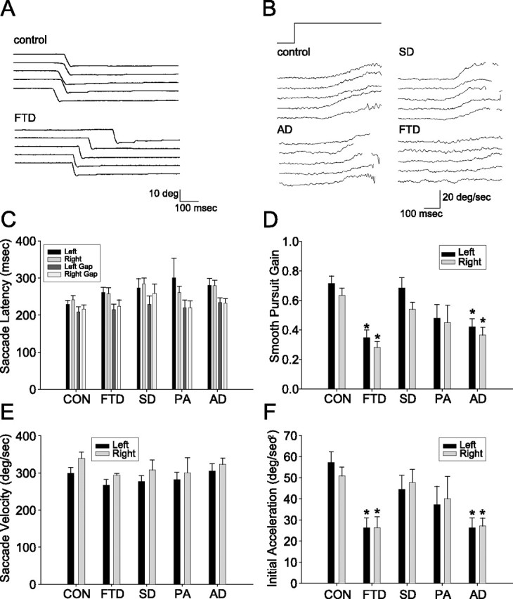
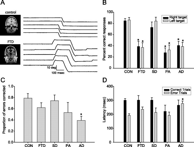
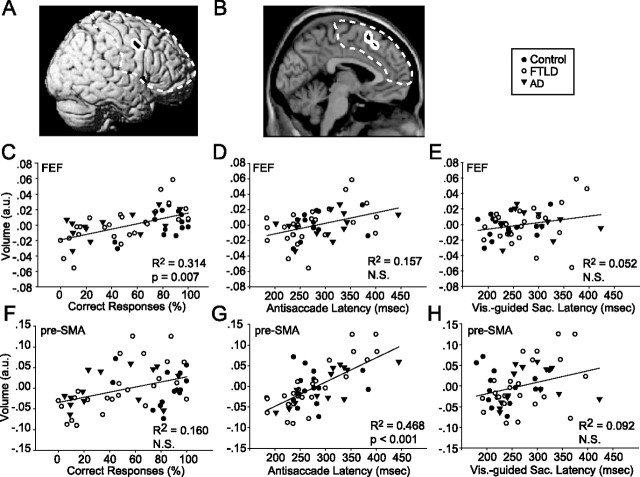
Deficits in the ability to suppress automatic behaviors lead to impaired decision making, aberrant motor behavior, and impaired social function in humans with frontal lobe neurodegeneration. We have studied patients with different patterns of frontal lobe dysfunction resulting from frontotemporal lobar degeneration or Alzheimer's disease, investigating their ability to perform visually guided saccades and smooth pursuit eye movements and to suppress visually guided saccades on the antisaccade task. Patients with clinical syndromes associated with dorsal frontal lobe damage had normal visually guided saccades but were impaired relative to other patients and control subjects in smooth pursuit eye movements and on the antisaccade task. The percentage of correct antisaccade responses was correlated with neuropsychological measures of frontal lobe function and with estimates of frontal lobe gray matter volume based on analyses of structural magnetic resonance images. After controlling for age, gender, cognitive status, and potential interactions between disease group and oculomotor function, an unbiased voxel-based morphometric analysis identified the volume of a segment of the right frontal eye field (FEF) as positively correlated with antisaccade performance (less volume equaled lower percentage of correct responses) but not with either pursuit performance or antisaccade or visually guided saccade latency or gain. In contrast, the volume of the presupplementary motor area (pre-SMA) and a portion of the supplementary eye fields correlated with antisaccade latency (less volume equaled shorter latency) but not with the percentage of correct responses. These results suggest that integrity of the presupplementary motion area/supplementary eye fields is critical for supervisory processes that slow the onset of saccades, facilitating voluntary saccade targeting decisions that rely on the FEF.


Figures

Figure 1.
Figure 1.
Visually guided saccades and smooth pursuit eye movements in FTLD and AD. A, Eye position traces showing examples of five successive downward saccades in a control subject and an FTD subject. B, Eye velocity traces showing examples of five consecutive pursuit responses from individual subjects from the control, AD, SD, and FTD groups. Target motion was 20 deg/s to the right. The rapid deflections of eye velocity associated with catch-up saccades have been removed. C–F, Bar graphs summarizing the oculomotor behavior of controls (CON) and all patient groups. In all graphs, black and gray bars show responses to rightward and leftward targets. Error bars show SDs across subject within each group. Asterisks indicate effects that were statistically significant relative to controls (p < 0.05, ANOVA, Tukey post hoc). C, Horizontal saccade latencies under overlap conditions and when a 200 ms gap is introduced between fixation offset and saccade onset. D, Mean gain of smooth pursuit eye movements. E, Mean velocity of horizontal saccades. F, Initial eye acceleration of horizontal smooth pursuit eye movements.
Figure 2.
Figure 2.
Antisaccade performance in FTLD and AD. A, Eye position records showing examples of five consecutive antisaccade trials in a 53-year-old female control and a 54-year-old female FTD subject. A correct antisaccade response would have been a downward deflection of the eye position trace. Note the initial errors followed by rapid corrections in all five traces for the FTD subject. The images at the left of the panel show coronal sections through the frontal cortex from T1-weighted MRI scans. B–D, Bar graphs summarizing antisaccade performance in controls (CON) and patient groups. In B and D, black and gray bars show responses to rightward and leftward targets. Error bars show SDs across subject within each group. Asterisks indicate effects that were statistically significant relative to controls (p < 0.05). B, Percentages of correct antisaccade trials. C, Antisaccade error correction rates. D. Antisaccade latency.
Figure 3.
Figure 3.
Relationship between regional gray matter volume and saccade parameters. A, B, Structural images of frontal lobe regions using black patches surrounded by a white border to indicate where gray matter volume is correlated (p < 0.1, corrected for multiple comparisons, family-wise error) with percentage of correct antisaccade responses (A) or antisaccade latency (B) from the VBM analysis. Dashed line indicates region on which statistics are based. C–F, Scatter plots showing the relationship between gray matter volume and oculomotor performance, where each symbol shows measurements from a different subject. Filled circles, open circles, and upside down triangles show data from control, FTLD, and AD subjects. Brain volume is expressed in arbitrary units from the VBM analysis at the most significantly correlated voxel in the regions displayed in A and B. C–E show data for the right FEF region indicated in A (MNI coordinates: x = 61, y = 4, z = 42). F–H show data for the pre-SMA region indicated in B (MNI coordinates: x = 0, y = 19, z = 58). C, F, Percentage of correct antisaccade responses. D, G, Antisaccade latencies combined across directions. E, H, Visually guided saccade latencies in the overlap condition.

References

    1. Abel LA, Unverzagt F, Yee RD (2002). Effects of stimulus predictability and interstimulus gap on saccades in Alzheimer's disease. Dement Geriatr Cogn Disord 13:235–243. - PubMed
    1. Ashburner J, Friston KJ (2000). Voxel-based morphometry–the methods. NeuroImage 11:805–821. - PubMed
    1. Bagary MS, Hutton SB, Symms MR, Barker GJ, Mutsatsa SH, Barnes TR, Joyce EM, Ron MA (2004). Structural neural networks subserving oculomotor function in first-episode schizophrenia. Biol Psychiatry 56:620–627. - PubMed
    1. Berman RA, Colby CL, Genovese CR, Voyvodic JT, Luna B, Thulborn KR, Sweeney JA (1999). Cortical networks subserving pursuit and saccadic eye movements in humans: an FMRI study. Hum Brain Mapp 8:209–225. - PMC - PubMed
    1. Blanke O, Seeck M (2003). Direction of saccadic and smooth eye movements induced by electrical stimulation of the human frontal eye field: effect of orbital position. Exp Brain Res 150:174–183. - PubMed

Publication types

MeSH terms