. 2006 Oct 1;108(7):2358-65.
doi: 10.1182/blood-2006-02-003475. Epub 2006 Jun 8.

Simultaneous activation of multiple signal transduction pathways confers poor prognosis in acute myelogenous leukemia

Affiliations

Simultaneous activation of multiple signal transduction pathways confers poor prognosis in acute myelogenous leukemia

Steven M Kornblau et al. Blood. .

Abstract

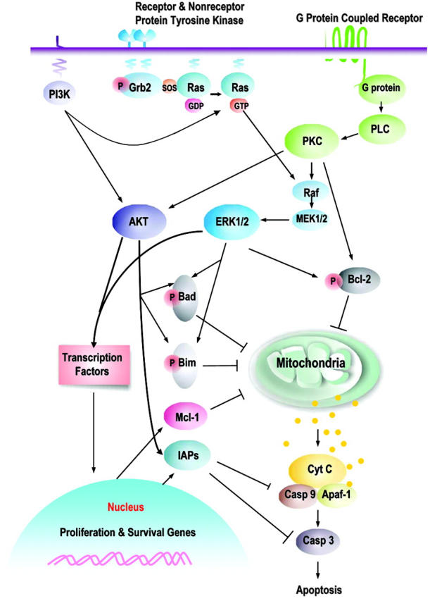
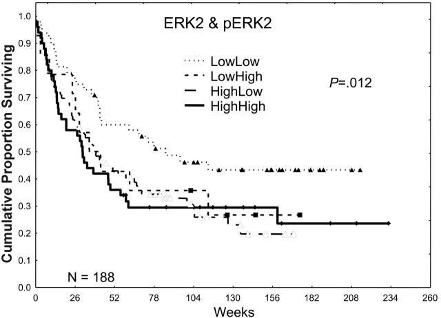
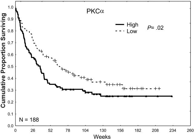
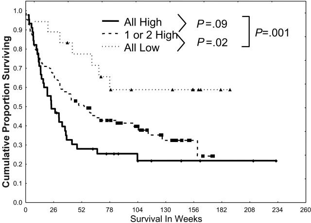
Deregulation of signal transduction pathways (STPs) may promote leukemogenesis by conferring cell proliferation and survival advantages in acute myelogenous leukemia (AML). Several agents targeting STPs are under development; however, redundancy and cross-talk between STPs could activate multiple downstream effectors and this could negate the effect of single-target inhibition. The frequency of concurrent activation of multiple STPs in AML and the prognostic relevance of STP activation in AML are unknown. STP protein expression (PKCalpha, ERK2, pERK2, AKT, and pAKT) was measured by Western blot in samples from 188 patients with newly diagnosed, untreated AML. In univariate and multivariate analysis high levels of PKCalpha, ERK, pERK, and pAKT, but not AKT, were adverse factors for survival as was the combination variable PKCalpha-ERK2&pERK2-pAKT. Survival progressively decreased as the number of activated pathways increased. Patients were more likely to have none or all 3 pathways activated than was predicted based on the frequency of individual pathway activation, strongly suggesting that cross-activation occurred. Simultaneous activation of multiple STPs is common in AML and has a progressively worse adverse effect on prognosis. It is thus likely that only combinations of agents that target the multiply activated STPs will be beneficial for patients with AML.


Figures

Figure 1.
Figure 1.
Prosurvival signaling pathways and their downstream targets. This is a schematic illustration of the principal pathways that are discussed in this paper. Growth factors induce, or membrane-associated molecules dimerize, spontaneously to activate mitogen-activated protein kinases (Raf/MEK/ERK) and PI3K/AKT signaling pathways. Transmembrane G protein–coupled receptors can activate PKC, which modulates the activity of Raf and MAPK pathway; both ERK1/2 and PKCα can serve as Bcl-2 kinases at serine 70. AKT and ERK induce phosphorylation of Bim (AKT, at serine 87 and ERK, at serines 55, 65, and 100), which attenuates the proapoptotic function of Bim, thereby promoting cell survival. Phosphorylation of Bad by MAPK (serine 112) or AKT (serine 136) inhibits apoptosis due to loss of the ability of Bad to heterodimerize with the survival proteins Bcl-2 and Bcl-XL. Hence, prosurvival phosphorylation of Bcl-2 family proteins modulates their antiapoptotic or proapoptotic activity at the mitochondrial membrane. This is complemented by AKT- or MAPK-driven gene transcription, which includes cyclins D and E (MAPK), cyclin-dependent kinase inhibitor p21, c-myc (AKT), which cause an increase in cell proliferation; and antiapoptotic proteins of the Bcl-2 (Mcl-1) and IAP (XIAP, survivin) families that regulate apoptosis at the level (Mcl-1) or downstream of mitochondria (IAPs). It is apparent that simultaneous activation of multiple signaling pathways might synergistically enhance prosurvival and proliferative potential of leukemic cells and the redundant downstream pathways negatively affect an ability of a particular signal transduction inhibitor to eliminate leukemia.
Figure 2.
Figure 2.
The effect of ERK2 and pERK2 expression on survival. Patients were divided into 2 groups on the basis of ERK2 expression, lower one third versus higher two thirds, on the basis of retrospective study data. Likewise, patients were divided into 2 groups, lower two thirds versus higher one third, on the basis of the level of pERK2 expression and retrospective study data. These were combined to form 4 groups for ERK2 and pERK2 (low/low, low/high, high/low, and high/high), and the Kaplan-Meier survival curves are shown here. The P value shown is for the plot of all 4 curves. P = .002 for the comparison of those with low/low ERK2 and pERK2 with those with one or both high.
Figure 3.
Figure 3.
The effect of PKCα expression on survival. Patients were divided into 2 groups, low and high, according to whether they were above or below the median PKCα expression observed in all 226 samples in 188 patients. Patients with a PKCα greater than the median level (P = .02) or in the highest third (P = .016) or the highest one sixth (P < .001) of this group had an inferior survival duration compared with those with levels of PKCα below the median, suggesting a progressively adverse prognostic effect of increasingly higher levels of PKCα.
Figure 4.
Figure 4.
The effect of pAKT expression on survival. Patients were divided into 2 groups, low and high, according to whether they were above or below the median pAKT expression observed in all 185 samples in 148 patients. Patients with high pAKT expression, greater than the median, had a survival duration inferior to that in patients with low pAKT expression, less than or equal to the median (P = .02).
Figure 5.
Figure 5.
Simultaneous activation of multiple STPs confers a poor prognosis. Patients were divided into 3 groups (all low, 1 or 2 high, or all 3 pathways highly activated) according to the activation of PKCα, ERK2 and pERK2, and pAKT and on the basis of the dichotomizations discussed in Figures 2, 3, 4. Patients were statistically more likely to have pan-activation or no activation than would be expected from the individual frequencies of low/high PKCα, ERK2 and pERK2, or pAKT. Survival was worse for those with 1 or 2 highly activated pathways (P = .02) and those with all 3 highly activated pathways (P < .001) than in those with all 3 pathways showing low levels of activation.

References

    1. Steelman LS, Pohnert SC, Shelton JG et al. JAK/STAT, Raf/MEK/ERK, PI3K/Akt and BCR-ABL in cell cycle progression and leukemogenesis. Leukemia. 2004;18: 189-218. - PubMed
    1. Reuter CW, Morgan MA, Bergmann L. Targeting the Ras signaling pathway: a rational, mechanism-based treatment for hematologic malignancies? Blood. 2000;96: 1655-1669. - PubMed
    1. Gilliland DG, Tallman MS. Focus on acute leukemias. Cancer Cell. 2002;1: 417-420. - PubMed
    1. Min YH, Cheong JW, Kim JY, et al. Cytoplasmic mislocalization of p27Kip1 protein is associated with constitutive phosphorylation of Akt or protein kinase B and poor prognosis in acute myelogenous leukemia. Cancer Res. 2004;64: 5225-5231. - PubMed
    1. Elstrom RL, Bauer DE, Buzzai M, et al. Akt stimulates aerobic glycolysis in cancer cells. Cancer Res. 2004;64: 3892-3899. - PubMed

Publication types