. 2006 Jul 12;25(13):3110-22.
doi: 10.1038/sj.emboj.7601187. Epub 2006 Jun 8.

Recruitment of PRC1 function at the initiation of X inactivation independent of PRC2 and silencing

Affiliations

Recruitment of PRC1 function at the initiation of X inactivation independent of PRC2 and silencing

Stefan Schoeftner et al. EMBO J. .

Abstract

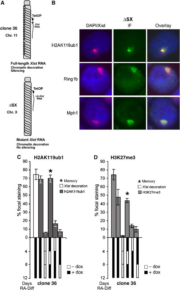
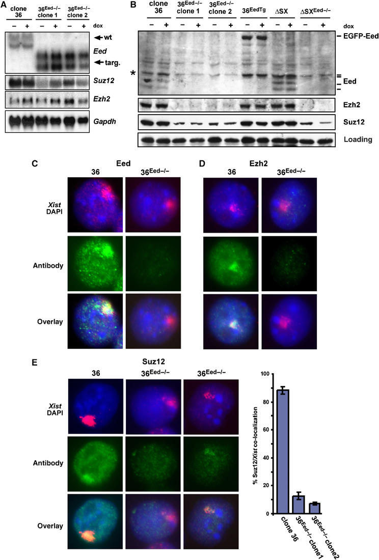
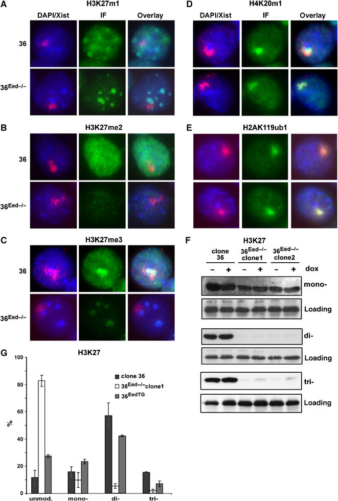
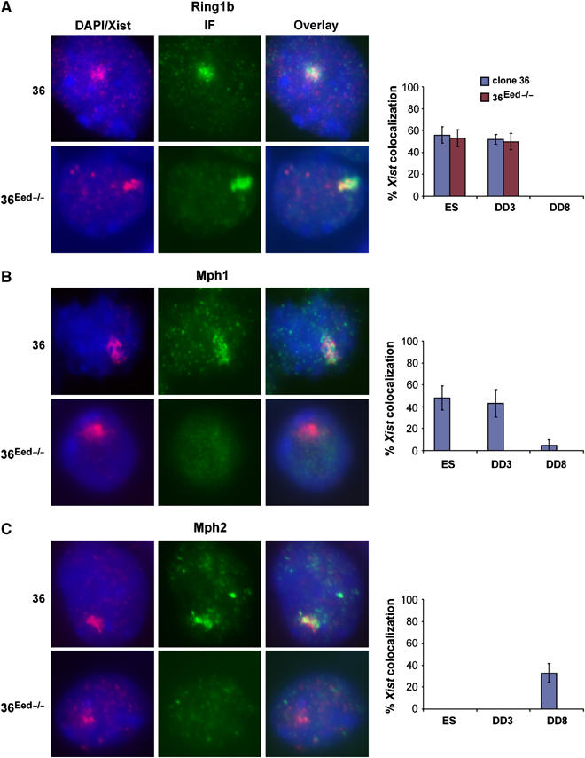
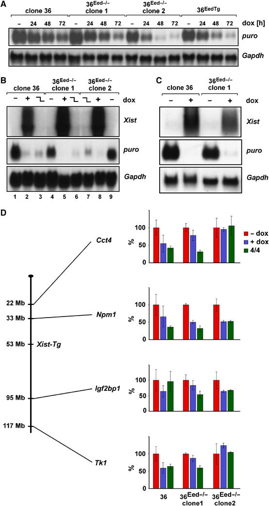
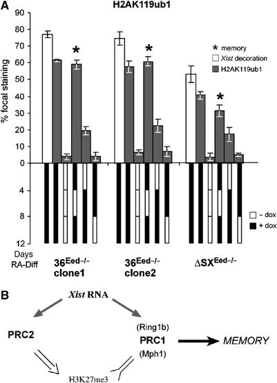
In mammals X inactivation is initiated by expression of Xist RNA and involves the recruitment of Polycomb repressive complex 1 (PRC1) and 2 (PRC2), which mediate chromosome-wide ubiquitination of histone H2A and methylation of histone H3, respectively. Here, we show that PRC1 recruitment by Xist RNA is independent of gene silencing. We find that Eed is required for the recruitment of the canonical PRC1 proteins Mph1 and Mph2 by Xist. However, functional Ring1b is recruited by Xist and mediates ubiquitination of histone H2A in Eed deficient embryonic stem (ES) cells, which lack histone H3 lysine 27 tri-methylation. Xist expression early in ES cell differentiation establishes a chromosomal memory, which allows efficient H2A ubiquitination in differentiated cells and is independent of silencing and PRC2. Our data show that Xist recruits PRC1 components by both PRC2 dependent and independent modes and in the absence of PRC2 function is sufficient for the establishment of Polycomb-based memory systems in X inactivation.


Figures

Figure 1
Figure 1
PRC1 recruitment by Xist. (A) Overview of the inducible Xist expression system (TetOP) on chromosome 11 and the X in clone 36 and ΔSX ES cells, respectively. In clone 36 ES cells, Xist induction silences a linked puromycin marker gene (puro). In ΔSX cells, the A repeat of Xist (triangle) is deleted. (B) Recruitment of the PRC1 components Ring1b and Mph1 as well as resulting H2AK119ub1 was observed by combined Xist RNA FISH (red) and immunofluorescence analysis (green) in undifferentiated ΔSX ES cells after 3 days Xist induction. (C) H2AK119ub1 is regulated by a chromosomal memory in differentiated cells. Bar graphs representing the percentage of nuclei with focal H2AK119ub1 signals (grey bars) and Xist RNA (white bars) is given (above). Error bars represent the standard deviation. Below a scheme of the ES cell differentiation time course showing the presence (black) or absence (white) of doxycycline. An asterisk marks the Xist induction scheme revealing the chromosomal memory. (D) Analysis of H3K27me3 in parallel cultures to (C).
Figure 2
Figure 2
Generation of ES cells lacking Eed. (A) Northern analysis of Eed, Suz12 and Ezh2 in undifferentiated control clone 36 and Eed deficient 36Eed-/- ES cells after Xist was induced for 3 days (+) or not (−); Gapdh as loading control. (B) Western analysis of Eed, Ezh2 and Suz12 in nuclear extracts from uninduced ES cells (−) or induced for 3 days (+). hnRNP A as loading control, asterisk indicates a nonspecific band. (C–E) Indirect immunofluorescence (green) of Eed (C), Ezh2 (D) or Suz12 (E) and subsequent Xist RNA FISH (red) of representative nuclei of undifferentiated 36Eed−/− and control clone 36 ES cells after 3 days of Xist induction. DAPI (blue) stains DNA. Statistics of the number of nuclei showing colocalisation of Suz12 staining with Xist in 36 and 36Eed−/− ES cells. Error bars indicate standard deviation (n>600).
Figure 3
Figure 3
Histone modifications in Eed deficient ES cells. (A–E) Combined Xist RNA FISH (red) indirect immunofluorescence of indicated histone modifications (green) analysis on undifferentiated 36Eed−/− and control 36 ES cells after 3 days Xist expression. Representative images are shown, statistics see Table I. (F) Western analysis of mono-, di- and tri-methylation of H3K27 in 36Eed−/− clones 1 and 2 and control 36 ES cells after Xist induction for 3 days (+) or not (−); loading control hnRNP A. (G) Mass-spectrometric analysis of histone H3 lysine 27 methylation in clone 36, 36Eed−/− and 36EedTG ES cells. The percentage of the indicated modification state is given for three independent experiments; error bars indicate standard deviation.
Figure 4
Figure 4
Recruitment of PRC1 components in the absence of Eed. (A, B) Indirect immunofluorescence (IF) of Ring1b (A), Mph1 (B) and subsequent Xist RNA FISH (red) analysis on undifferentiated 36Eed−/− and control clone 36 ES cells after Xist expression for 3 days. (C) Analysis for Mph2 in ES cells differentiated for 8 days in the presence of doxycycline. The percentage of nuclei showing focal IF staining colocalising with Xist RNA is given for undifferentiated (ES), day 3 (DD3) and day 8 (DD8) of differentiation. Error bars represent standard deviation (n>350).
Figure 5
Figure 5
Initiation and maintenance of silencing independent of Eed. (A) Northern analysis of PGKpuromycin, (puro) silencing in 36Eed−/− and control clone 36 cells after Xist induction for 24, 48 and 72 h; Gapdh as loading control. (B) Maintenance of puro silencing in cells differentiated in the presence (+; lanes 2, 5 and 8) or absence (−; 1, 4 and 9) doxycycline, or differentiated for 4 days in the presence followed by 4 days in the absence of doxycycline (lanes 3, 6 and 7). (C) Northern analysis of puro expression in embryoid bodies outgrowths established in the presence of doxycycline (+) or without (−) after 4 weeks. (D) Quantitative expression analysis of Cct4, Npm1, Igf2b and Tk1 on chromosome 11 in control 36 and 36Eed-/− ES cells at day 8 of differentiation in the absence (red bars), continuous presence (blue bars) of doxycycline, or presence of doxycycline for the first 4 days (green bars). Means of three independent measurements normalised to Gapdh are shown, error bars represent standard deviation. Scheme on the left shows the genes relative to the Xist transgene.
Figure 6
Figure 6
A chromosomal memory independent of silencing and Eed. (A) The percentage of nuclei showing focal staining for H2AK119ub1 or H3K27me3 (grey bars) and Xist RNA (white bars) is given (above). Error bars represent the standard deviation. Underneath the ES cell differentiation time course is depicted and the presence (black) or absence (white) of doxycycline is indicated. The Xist induction scheme revealing a chromosomal memory for H2AK119ub1 and H3K27me3 is marked by an asterisk. (B) Summary of recruitment of PRC1 and PRC2 by Xist in X inactivation.

References

    1. Atsuta T, Fujimura S, Moriya H, Vidal M, Akasaka T, Koseki H (2001) Production of monoclonal antibodies against mammalian Ring1B proteins. Hybridoma 20: 43–46 - PubMed
    1. Birve A, Sengupta AK, Beuchle D, Larsson J, Kennison JA, Rasmuson-Lestander A, Muller J (2001) Su(z)12, a novel Drosophila Polycomb group gene that is conserved in vertebrates and plants. Development 128: 3371–3379 - PubMed
    1. Borsani G, Tonlorenzi R, Simmler MC, Dandolo L, Arnaud D, Capra V, Grompe M, Pizzuti A, Muzny D, Lawrence C et al. (1991) Characterization of a murine gene expressed from the inactive X chromosome. Nature 351: 325–329 - PubMed
    1. Brockdorff N, Ashworth A, Kay GF, Cooper P, Smith S, McCabe VM, Norris DP, Penny GD, Patel D, Rastan S (1991) Conservation of position and exclusive expression of mouse Xist from the inactive X chromosome. Nature 351: 329–331 - PubMed
    1. Brown CJ, Ballabio A, Rupert JL, Lafreniere RG, Grompe M, Tonlorenzi R, Willard HF (1991a) A gene from the region of the human X inactivation centre is expressed exclusively from the inactive X chromosome. Nature 349: 38–44 - PubMed

Publication types

MeSH terms