. 2006 Jun 21;25(12):2847-55.
doi: 10.1038/sj.emboj.7601178. Epub 2006 Jun 8.

Controlling the subcellular localization of DNA polymerases iota and eta via interactions with ubiquitin

Affiliations

Controlling the subcellular localization of DNA polymerases iota and eta via interactions with ubiquitin

Brian S Plosky et al. EMBO J. .

Abstract

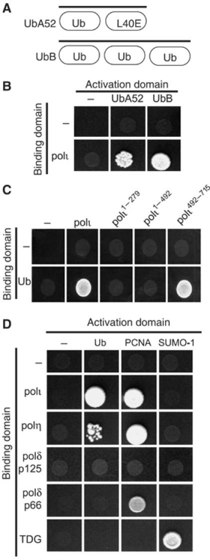
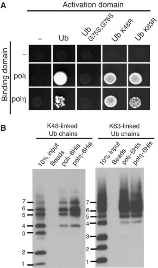
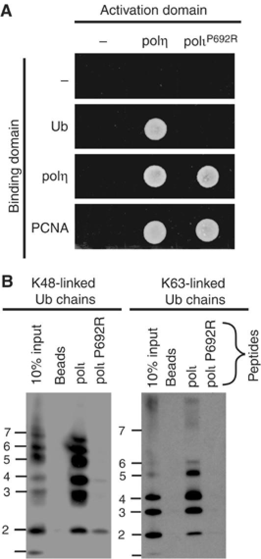
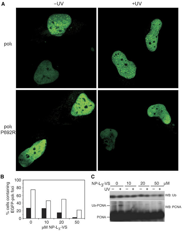
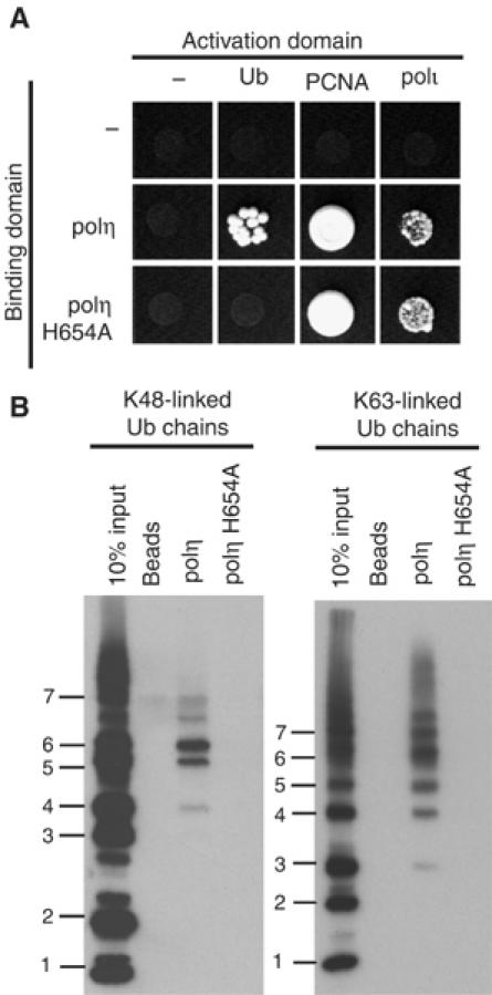
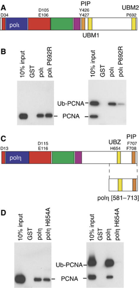
Y-family DNA polymerases have spacious active sites that can accommodate a wide variety of geometric distortions. As a consequence, they are considerably more error-prone than high-fidelity replicases. It is hardly surprising, therefore, that the in vivo activity of these polymerases is tightly regulated, so as to minimize their inadvertent access to primer-termini. We report here that one such mechanism employed by human cells relies on a specific and direct interaction between DNA polymerases iota and eta with ubiquitin (Ub). Indeed, we show that both polymerases interact noncovalently with free polyUb chains, as well as mono-ubiquitinated proliferating cell nuclear antigen (Ub-PCNA). Mutants of poliota (P692R) and poleta (H654A) were isolated that are defective in their interactions with polyUb and Ub-PCNA, whilst retaining their ability to interact with unmodified PCNA. Interestingly, the polymerase mutants exhibit significantly lower levels of replication foci in response to DNA damage, thereby highlighting the biological importance of the polymerase-Ub interaction in regulating the access of the TLS polymerases to stalled replication forks in vivo.


Figures

Figure 1
Figure 1
Yeast two-hybrid interactions between TLS polymerases and Ub. (A) Diagram of the gene structure of UbA52 and UbB. (B) Yeast two-hybrid assay showing the interaction (by activation of the ADE2 and HIS3 reporter genes) between full-length polι and UbA52 and UbB. In these, and all subsequent two-hybrid assays, 10 μl of an overnight culture was spotted on CSM -Leu, -Trp, -His, -Ade plates and incubated at 30°C for 5 days. (C) Deletion mapping of polι reveals that the interaction with recombinant Ub is localized to the C-terminal 224 amino acids of polι. (D) In addition to polι, polη also binds to Ub. However, neither the p125 and p66 subunits of polδ bind Ub. PCNA was used as a positive control for polι, polη, and p66 of polδ. The interaction between polι and polη and Ub is specific, as neither polymerase binds to the Ub-like protein, SUMO-1. TDG was used as a positive control for SUMO-1.
Figure 2
Figure 2
The interaction between pols ι and η and Ub is noncovalent, but requires Ub conjugation. (A) While polι and polη interact with wild-type Ub, neither interacts with the G75S/G76S Ub mutant that cannot be conjugated to a target protein or form polyUb chains. Additionally, polι and polη are able to interact with either K48R- or K63R-Ub. (B) In vitro pull down assays with purified C-terminal His-tagged polι and polη demonstrate that both polymerases interact with pure K48- and K63-linked polyUb chains ranging from four to seven Ub moieties. Bound proteins were separated on 10–20% Tris–glycine–SDS–polyacrylamide gels and transferred onto PVDF membrane and Ub chains visualized by Western blot.
Figure 3
Figure 3
Identification of a mutant polι that cannot bind Ub. (A) polι P692R has lost the ability to bind Ub, but can still bind polη and PCNA in the yeast two-hybrid assay. (B) Pull-down assay using biotinylated peptides corresponding to the C-terminus of wild-type polι or a P692R variant and either K48- or K63-linked polyUb chains were analyzed as described in Figure 2B. Only the peptide corresponding to the wild-type polι sequence is able to bind either form of polyUb chain.
Figure 4
Figure 4
Binding to Ub is required for polι to relocalize into replication foci. (A) Wild-type pEGFP polι (upper panels) or pEGFP polι P692R (lower panels) were transfected into MRC5 cells and either mock treated (left hand panels) or irradiated with 7 J/m2 UV (right-hand panels). Nuclei from two adjacent cells are shown in each panel for comparison. (B) Histogram showing the percentage of nuclei with foci after exposure to various concentrations of the 26S proteasome inhibitor, NP-L3-VS before (closed bars) and after UV-irradiation (open bars). Numbers are based upon 200 nuclei analyzed per data point. (C) Western blot of Ub or PCNA in whole-cell extracts (50 μg) separated by SDS–PAGE on a 15% gel, after treatment with NP-L3-VS with and without UV-irradiation (7 J/m2). Note that the levels of Ub and Ub-PCNA decrease as the dose of NP-L3-VS is increased.
Figure 5
Figure 5
A mutant in polη's Zn-finger motif is defective in binding Ub. (A) Yeast two-hybrid assays showing that the polη H654A mutant is unable to interact with Ub, but nevertheless retains its ability to interact with full-length polι and PCNA. (B) Full-length C-terminal His-tagged polη or polη-H654A were used in pull-down assays with K48- or K63-linked Ub chains demonstrating that the H654A mutant is defective at binding either form of polyUb. Bound proteins were analyzed as described in Figure 2B.
Figure 6
Figure 6
Protein–protein interactions regulating TLS. (A) Schematic representation of the domain/motif structure of polι. The polymerase domain representing conserved finger (blue) palm (red), thumb (green) and little finger (purple) spans the N-terminal portion of each protein. The PCNA binding site (PIP-Box) of each protein is colored orange and the UBMs are in yellow. UBM1 spans residues 496–524 and UBM2 spans amino acids 681–709 (Bienko et al, 2005). Residues identified in the active site of the polymerase, PIP-box or Ub interactions are shown by the single letter code. (B) Full-length wild-type GST-polι or polι-P692R mutant were used in pull-down assays with either PCNA or ubiquitinated-PCNA, separated on 10–20% Tris–glycine–SDS gels, transferred onto PVDF membrane, and PCNA was detected by Western Blot. Left: Both wild-type, and P692R polι GST-fusion proteins are able to bind to PCNA. Right: However, polι-P692R has a reduced ability to pull down Ub-PCNA. (C) Schematic representation of the domain/motif structure of polη (same color scheme as (A)), with amino acids 631–659 comprising the UBZ domain (Bienko et al, 2005). (D) Pull-down assays as described above were carried out with GST fused to the C-terminal 133 amino acids of polη or polη-H654A. Left: GST-polη[581–713] contains the PIP box and both wild-type polη, or the H654A polη mutant can bind PCNA, in accord with data shown in Figure 4B. Right: However, while the wild-type polη protein is able to pull-down Ub-PCNA, the H654A polη-Ub mutant is unable to bind ubiquitinated PCNA.

References

    1. Baba D, Maita N, Jee JG, Uchimura Y, Saitoh H, Sugasawa K, Hanaoka F, Tochio H, Hiroaki H, Shirakawa M (2005) Crystal structure of thymine DNA glycosylase conjugated to SUMO-1. Nature 435: 979–982 - PubMed
    1. Bailly V, Lamb J, Sung P, Prakash S, Prakash L (1994) Specific complex formation between yeast RAD6 and RAD18 proteins: a potential mechanism for targeting RAD6 ubiquitin-conjugating activity to DNA damage sites. Genes Dev 8: 811–820 - PubMed
    1. Bailly V, Lauder S, Prakash S, Prakash L (1997) Yeast DNA repair proteins Rad6 and Rad18 form a heterodimer that has ubiquitin conjugating, DNA binding, and ATP hydrolytic activities. J Biol Chem 272: 23360–23365 - PubMed
    1. Bienko M, Green CM, Crosetto N, Rudolf F, Zapart G, Coull B, Kannouche P, Wider G, Peter M, Lehmann AR, Hofmann K, Dikic I (2005) Novel ubiquitin-binding domains in Y-family polymerases regulate translesion DNA synthesis. Science 310: 1821–1824 - PubMed
    1. Chau V, Tobias JW, Bachmair A, Marriott D, Ecker DJ, Gonda DK, Varshavsky A (1989) A multiubiquitin chain is confined to specific lysine in a targeted short-lived protein. Science 243: 1576–1583 - PubMed

Publication types

MeSH terms