. 2006 Jun 21;25(12):2710-22.
doi: 10.1038/sj.emboj.7601169. Epub 2006 Jun 8.

Roles for APIS and the 20S proteasome in adenovirus E1A-dependent transcription

Affiliations

Roles for APIS and the 20S proteasome in adenovirus E1A-dependent transcription

Mozhgan Rasti et al. EMBO J. .

Abstract

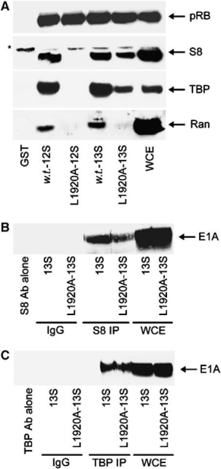
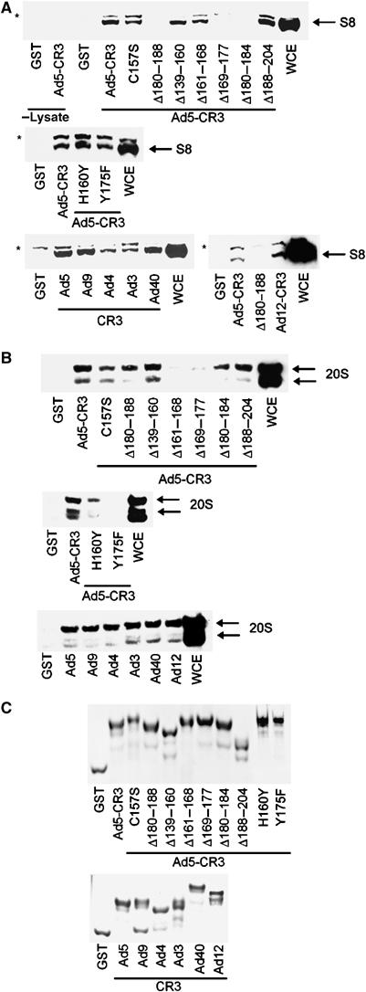
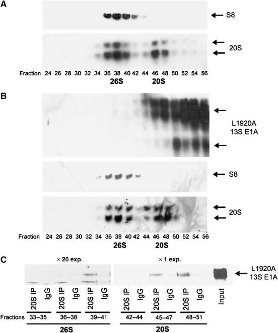
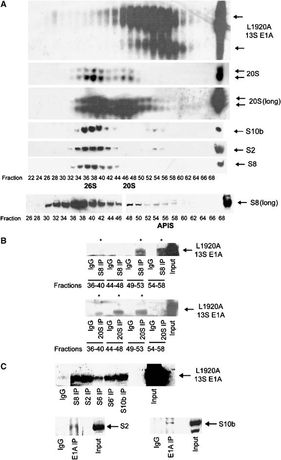
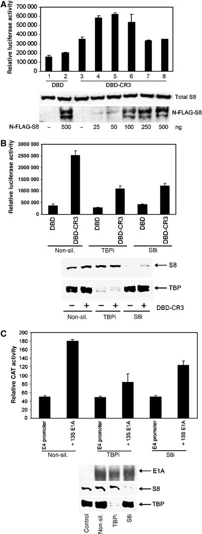
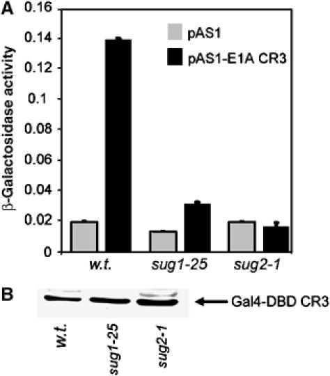
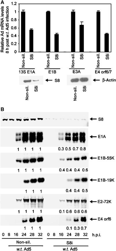
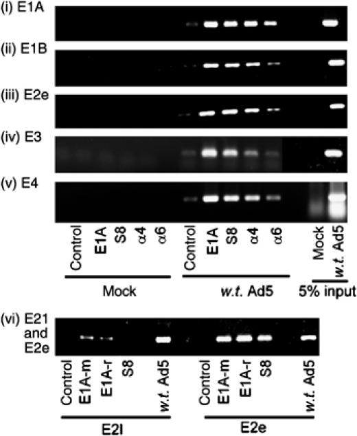
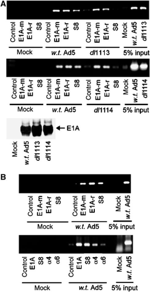
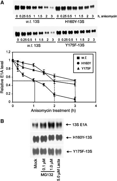
We have determined distinct roles for different proteasome complexes in adenovirus (Ad) E1A-dependent transcription. We show that the 19S ATPase, S8, as a component of 19S ATPase proteins independent of 20S (APIS), binds specifically to the E1A transactivation domain, conserved region 3 (CR3). Recruitment of APIS to CR3 enhances the ability of E1A to stimulate transcription from viral early gene promoters during Ad infection of human cells. The ability of CR3 to stimulate transcription in yeast is similarly dependent on the functional integrity of yeast APIS components, Sug1 and Sug2. The 20S proteasome is also recruited to CR3 independently of APIS and the 26S proteasome. Chromatin immunoprecipitation reveals that E1A, S8 and the 20S proteasome are recruited to both Ad early region gene promoters and early region gene sequences during Ad infection, suggesting their requirement in both transcriptional initiation and elongation. We also demonstrate that E1A CR3 transactivation and degradation sequences functionally overlap and that proteasome inhibitors repress E1A transcription. Taken together, these data demonstrate distinct roles for APIS and the 20S proteasome in E1A-dependent transactivation.


Figures

Figure 1
Figure 1
S8 binds to CR3 of Ad5 13S E1A. (A) GST pull-downs using w.t. 12S and 13S E1A, and 12S L1920A and 13S L1920A E1A mutants, were performed to assess the in vitro binding capacity of these E1A proteins for pRB, S8, TBP and Ran from A549 cellular lysates. Following pull-down, binding was assessed by Western blotting. (B, C) S8 and TBP retain the ability to bind L1920A 13S E1A in vivo. S8 and TBP complexes were immunoprecipitated from 13S E1A-, and 13S E1A L1920A-, expressing A549 cells, and subjected to Western blotting for E1A. *Nonspecific band recognised by the anti-S8 antibody. WCE, whole-cell extract.
Figure 2
Figure 2
Binding of S8 and 20S proteasomal components to Ad5 CR3 E1A mutants, and to CR3s from different Ad subgroups. Following GST pull-down, binding capacity was assessed by Western blotting for S8 (A) and 20S proteasomes (B). The 20S proteasome antibody recognises α1–α3 and α5–α7 subunits. In lanes 1 and 2 of the upper panel of (A), GST and GST-Ad5 CR3 were incubated in lysis buffer alone. (C) Coomassie-stained gel showing the purity of the E1A proteins used. *Nonspecific band recognised by the anti-S8 antibody. WCE, whole-cell extract. The corresponding regions of CR3 used during this study were as follows: Ad5, residues 139–204; Ad3, residues 132–209; Ad4, residues 128–206; Ad9, residues 120–198; Ad12, residues 124–210; Ad40, residues 117–193.
Figure 3
Figure 3
Binding of 20S proteasomes to 13S L1920A in vivo. A549 (A) and 13S-L1920A A549 (B) cellular lysates were fractionated upon a Superose 6 HR 10/30 column by FPLC (Materials and methods). E1A, and 26S, and 20S, proteasome-containing fractions were identified by Western blotting. Fractions containing 26S proteasomes or 20S proteasomes were pooled, and immunoprecipitation was performed either with an antibody that immunoprecipitates 20S proteasomal components or a normal IgG for control immunoprecipitations. Immunoprecipitates were separated upon a urea gel, and E1A identified by Western blotting (C). The blots indicate that L1920A 13S E1A binds significantly better to bulk 20S proteasomes (× 1 exposure) than 20S proteasomes associated with 26S proteasomes (× 20 exposure).
Figure 4
Figure 4
Binding of S8 as a component of APIS to 13S L1920A in vivo. (A) 13S-L1920A A549 cellular lysates were fractionated by FPLC and E1A-, 26S-, 20S- and APIS-containing fractions identified by Western blotting. Long exposures (long) were performed to identify all fractions containing 26S, 20S and APIS. Fractions comprising 26S proteasomes, or 20S proteasomes or APIS were pooled and immunoprecipitation was performed either with an antibody that immunoprecipitates S8 (B, upper panel), an antibody that immunoprecipitates 20S proteasomal components (B, lower panel) or a normal IgG for control immunoprecipitations (B). Immunoprecipitates were separated upon a urea gel, and E1A identified by Western blotting (B). The blots indicate that L1920A 13S E1A binds significantly better to S8 through APIS than through 26S proteasomes (B, upper panel), and that L1920A 13S E1A binds significantly better to bulk 20S proteasomes rather than 20S proteasomes associated with 26S (B, lower panel). L1920A 13S E1A also associates with other 19S base (APIS) components in vivo (C). S8, S2, S6, S6′ and S10b were immunoprecipitated from 13S-L1920A A549 cellular lysates and immunoprecipitated E1A* identified by Western blotting.
Figure 5
Figure 5
Requirement for S8 and TBP in 13S E1A transactivation function. (A) Effect of S8 expression upon E1A transactivation function. HCT116 cells were transfected with a constant amount of pcDNA3-Gal4DBD-CR3, and a Gal4-responsive luciferase reporter, with increasing amounts of pcDNA3-N-FLAG-S8. At 24 h post-transfection, cell lysates were prepared and luciferase activities measured. Total S8 and N-FLAG S8 were quantified by Western blotting. (B) Effect of TBP and S8 knock-down upon E1A transactivation. Following knock-down, HCT116 cells were transfected with a constant amount of pcDNA3-Gal4DBD and a Gal4-responsive luciferase reporter or pcDNA3-Gal4DBD-CR3 and a Gal4-responsive luciferase reporter. At 24 h post-transfection, cell lysates were prepared and luciferase activities measured. S8 and TBP levels were determined by Western blotting. (C) Following knock-down, HCT116 cells were transfected with an Ad5 E4 promoter-tethered CAT reporter and pcDNA3 Ad5 13S E1A. At 24 h post-transfection, cell lysates were prepared and CAT activities measured. In all instances, transfected DNA levels were equalised using pcDNA3 alone. Results are the mean±s.d. from three independent experiments. E1A, S8 and TBP levels were determined by Western blotting. The ability of pLE2 to transactivate the Ad5 E4 promoter was also inhibited significantly following knock-down of TBP or S8 (data not shown).
Figure 6
Figure 6
A requirement for Sug1 and Sug2 in CR3 transactivation function in yeast. W.t., sug1-25 and sug2-1 yeast strains were transformed with a Gal4-reporter construct and Gal4DBD or Gal4DBD-CR3. Cell lysates were subsequently prepared and β-galactosidase assays (A) performed to determine the transactivation capacity of CR3 in mutant sug1-25, mutant sug2-1 and the congenic w.t. strain. Western blotting analyses were also performed (B) with an anti-GAL4DBD antibody to determine the levels of E1A protein expression. Results are the mean±s.d. from three independent experiments.
Figure 7
Figure 7
The effect of S8 knock-down upon Ad early region gene, and gene product, expression during Ad infection. Following knock-down, A549 cells were infected with w.t. Ad5 at 1 PFU/cell. At the indicated time post-infection, total RNA was prepared and the absolute levels of 13SE1A, the 2.2 kb E1B transcript, E3A and E4 orf6/7 mRNAs were determined (A) by quantitative real-time PCR. In each instance, Ad early mRNA levels in non-silencing controls were ascribed a value of 100% expression. mRNA levels in knock-down cells were calculated on this basis. Values are the mean±s.d. from three independent experiments. At the appropriate time post-infection, cell lysates were prepared and the relative levels of S8, E1A, E1B-55K, E1B-19K, E2-72K and E4ORF6 proteins were determined (B) by Western blotting.
Figure 8
Figure 8
E1A, S8 and 20S proteasomal components are recruited to Ad early promoters in vivo during a w.t. Ad5 infection. Following infection, E1A, S8 and 20S protein–DNA complexes were isolated by ChIP. Typically, 10 μg of each antibody was used for ChIP. A normal rabbit IgG was used as a control antibody. E1A, E1B, E2e, E3, E4 and E2l promoter elements were detected by PCR. E1A-m, mouse monoclonal; E1A-r, rabbit polyclonal. Similar results were obtained from three independent experiments.
Figure 9
Figure 9
(A) Recruitment of S8 to Ad early region promoters is dependent upon binding to CR3. A549 cells were infected with either w.t. Ad5, or Ad5 mutants, dl1113 (panel 1) or dl1114 (panel 2), at an m.o.i. of 1 PFU/cell. At the appropriate time post-infection, E1A and S8 protein–DNA complexes were isolated by ChIP and E3 promoter elements detected by PCR. E1A expression was determined by Western blotting in the lower panel. (B) A role for E1A and S8 in transcriptional elongation. Following infection with w.t. Ad5, E1A, S8 and 20S protein–DNA complexes were isolated by ChIP and E1B gene elements detected by PCR. Upper panel, nucleotides 1751–2030 were amplified by PCR; lower panel, nucleotides 2591–2930 were amplified by PCR; typically 10 μg of each antibody was used for ChIP. A normal rabbit IgG was used as a control antibody. E1A-m, mouse monoclonal; E1A-r, rabbit polyclonal. Similar results were obtained from three independent experiments.
Figure 10
Figure 10
The protein half-lives of transactivation-defective 13S E1A mutants are extended relative to w.t. 13S E1A (A). Stably expressing 13S E1A-A549 cells, 13S H160Y E1A-A549 cells and 13S Y175F E1A-A549 cells were treated with the protein synthesis inhibitor anisomycin (100 μM) and harvested at the indicated times. E1A levels were determined by Western blotting and quantified using a Bio-Rad GS-800 densitometer. Results shown are the mean±s.d. from four independent experiments. The half-lives of 13S H160Y E1A and 13S Y175F E1A were similarly enhanced relative to w.t. 13S E1A when assessed by Western blotting (with E1A mAb, M2) of anisomycin-treated A549 cells transiently transfected with pLE2-expressing w.t. or mutant 13S E1A proteins (data not shown). Sensitivity of w.t. 13S E1A, H160Y-13S and Y175F-13S protein levels to treatment with proteasome inhibitors (B). Stably expressing 13S E1A-A549 cells, 13S H160Y E1A-A549 cells and 13S Y175F E1A-A549 cells were treated with the indicated amounts of proteasome inhibitors for 8 h. E1A levels were determined by Western blotting. Proteasome inhibitors also enhanced w.t. 13S E1A protein levels in A549 cells when transiently expressed using pLE2, but were unable to enhance H160Y E1A and 13S Y175F E1A protein levels when similarly expressed transiently using pLE2 (data not shown).

References

    1. Avvakumov N, Wheeler R, D'Halluin JC, Mymryk JS (2002) Comparative sequence analysis of the largest E1A proteins of human and simian adenoviruses. J Virol 76: 7968–7975 - PMC - PubMed
    1. Boyer TG, Berk AJ (1993) Functional interaction of adenovirus E1A with holo-TFIID. Genes Dev 7: 1810–1823 - PubMed
    1. Boyer TG, Martin ME, Lees E, Ricciardi RP, Berk AJ (1999) Mammalian Srb/Mediator complex is targeted by adenovirus E1A protein. Nature 399: 276–279 - PubMed
    1. Chiang CM, Roeder RG (1995) Cloning of an intrinsic human TFIID subunit that interacts with multiple transcriptional activators. Science 267: 531–536 - PubMed
    1. Ciechanover A (1998) The ubiquitin–proteasome pathway: on protein death and cell life. EMBO J 17: 7151–7160 - PMC - PubMed

Publication types

Substances