. 2006 Jun 21;25(12):2792-801.
doi: 10.1038/sj.emboj.7601159. Epub 2006 Jun 8.

Musashi regulates the temporal order of mRNA translation during Xenopus oocyte maturation

Affiliations

Musashi regulates the temporal order of mRNA translation during Xenopus oocyte maturation

Amanda Charlesworth et al. EMBO J. .

Abstract

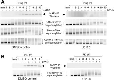
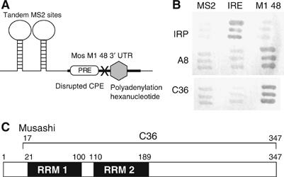
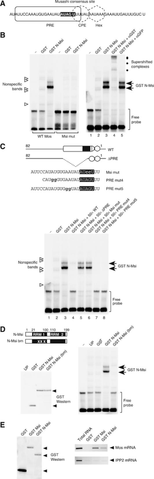
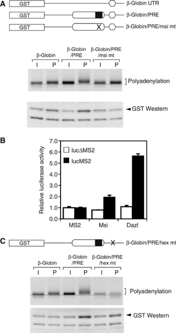
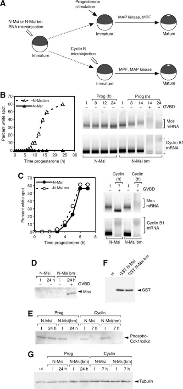
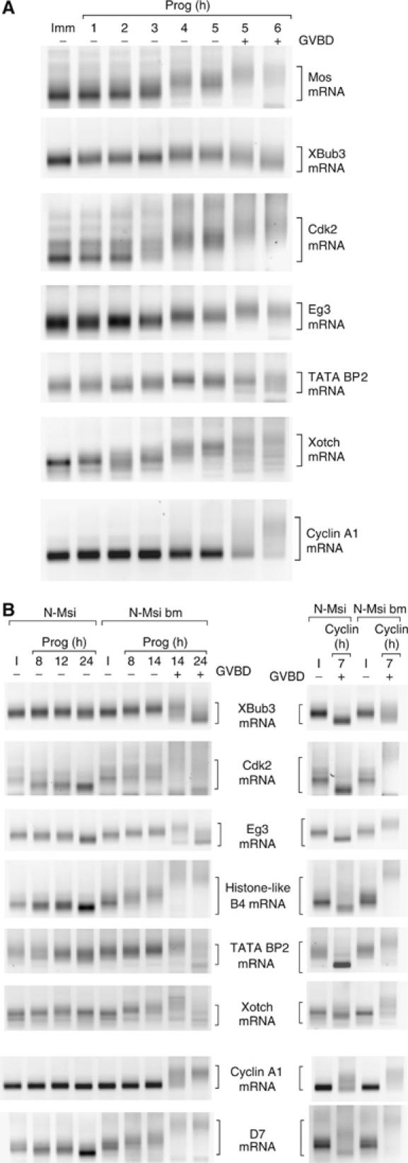
A strict temporal order of maternal mRNA translation is essential for meiotic cell cycle progression in oocytes of the frog Xenopus laevis. The molecular mechanisms controlling the ordered pattern of mRNA translational activation have not been elucidated. We report a novel role for the neural stem cell regulatory protein, Musashi, in controlling the translational activation of the mRNA encoding the Mos proto-oncogene during meiotic cell cycle progression. We demonstrate that Musashi interacts specifically with the polyadenylation response element in the 3' untranslated region of the Mos mRNA and that this interaction is necessary for early Mos mRNA translational activation. A dominant inhibitory form of Musashi blocks maternal mRNA cytoplasmic polyadenylation and meiotic cell cycle progression. Our data suggest that Musashi is a target of the initiating progesterone signaling pathway and reveal that late cytoplasmic polyadenylation element-directed mRNA translation requires early, Musashi-dependent mRNA translation. These findings indicate that Musashi function is necessary to establish the temporal order of maternal mRNA translation during Xenopus meiotic cell cycle progression.


Figures

Figure 1
Figure 1
The Mos PRE is responsive to a MAP kinase-independent trigger pathway. (A) Immature oocytes were injected with RNA encoding the GST coding region fused to the β-globin 3′ UTR containing the Mos PRE (β-globin/PRE). Oocytes were pretreated where indicated with U0126 and then stimulated with progesterone. Protein and RNA were extracted from the same pools of oocytes at the specified times. Progression of maturation is indicated by whether the oocytes had (+) or had not (−) undergone GVBD. Top panel is a Western blot for active (phosphorylated) MAP kinase (arrowhead). Lysates from DMSO- and UO126-treated oocytes were processed and analyzed in parallel. Lower panels show polyadenylation (brackets) of the endogenous Mos and cyclin B1 mRNAs and the injected synthetic β-globin/PRE reporter mRNA, as assayed by RNA ligation-coupled RT–PCR from the same cDNA preparation. Retardation of the PCR products is indicative of polyadenylation. (B) Immature oocytes were injected with the β-globin/PRE reporter. Oocytes were pretreated where indicated with U0126 and then stimulated by injection of recombinant PKI protein. Top panel is a Western blot for active (phosphorylated) MAP kinase (arrowhead). Lysates from DMSO-and UO126-treated oocytes were processed and analyzed in parallel. Lower panels show polyadenylation of the injected synthetic β-globin/PRE reporter (brackets).
Figure 2
Figure 2
Identification of Mos UTR-specific binding proteins by yeast three-hybrid analyses. (A) Diagram of the RNA hybrid in the pIIIA/MS2 vector that was used to screen the Xenopus oocyte library. The last 48 nt of the Mos 3′ UTR (M1 48, bold line) containing the PRE (open rectangle), the canonical polyadenylation hexanucleotide (gray hexagon) and a disrupted CPE (‘X', UUUUAU to UUUggU) were placed 3′ of the MS2 sites. (B) Clones A8 and C36 specifically bind to the Mos M1 48 hybrid RNA. pACT2 plasmids encoding A8 and C36 clones and IRP (iron response protein) hybrid proteins were co-transformed with pIIIA/MS2 plasmids specifying MS2 (empty vector), IRE/MS2 (IRE: iron response element) and MS2/M1 48 (M1 48) hybrid RNAs. Dark gray shows activation of the LacZ reporter, indicating interaction between protein and RNA. (C) Schematic of the Xenopus Musashi protein. The black boxes represent the RNA recognition motifs (RRM) of Musashi. Amino-acid position of the motifs and the fragment of Musashi (C36) that was recovered from the screen are indicated.
Figure 3
Figure 3
Musashi binds to the PRE in the Mos 3′ UTR. (A) Schematic showing regulatory element composition within the last 50 nt of the Mos 3′ UTR. The Musashi consensus binding site is indicated by white nucleotides. (B) Left panel: Wild-type (WT Mos) or Musashi binding site mutant (Msi mut) Mos UTR probes were analyzed for interaction with the GST-tagged, N-terminal domain of Xenopus Musashi (GST N-Msi) by RNA EMSA. Specific Musashi binding complexes were only detected with the wild-type Mos UTR probe. Right panel: Musashi-specific complex formation with the wild-type Mos UTR probe can be supershifted with antisera to the GST epitope tag and not with GFP antisera. (C) Musashi-specific complex formation with the wild-type Mos UTR probe can be competed with a 50-fold excess unlabeled wild-type, PRE mut4 and PRE mut5 Mos UTR RNA. No competition is observed with a Mos UTR lacking the entire PRE (ΔPRE) or a Musashi binding site mutant (Msi mut) Mos UTR. (D) Disruption of the first RRM in Musashi (GST N-Msi-bm) prevents Musashi-specific complex formation with the wild-type Mos UTR probe (right panel). Equivalent levels of each GST fusion protein were expressed in the reticulocyte lysates (left panel). UP, unprogrammed lysate. (E) The full-length (GST Msi) and N-terminal domain of Musashi (GST N-Msi) interact with the endogenous Mos mRNA. Left panel: GST Western blot showing recovery of exogenously expressed GST fusion proteins using glutathione Sepharose beads. Equivalent levels of GST Msi and GST N-Msi and higher levels of the GST moiety were recovered in the pulldown. Right panel: RT–PCR of Mos and IPP2 mRNA from RNA extracted from the indicated GST fusion proteins shown in the left panel. GST-Msi and GST-N-Msi interact with the Mos mRNA but do not interact with the IPP2 mRNA. The GST moiety alone fails to interact with either Mos or IPP2 mRNAs. RT–PCR from total oocyte RNA was used as a positive control to indicate the relative position of the expected PCR products in the pulldown lanes.
Figure 4
Figure 4
Musashi directs mRNA translational activation. (A) Upper panel: Schematic representation of the reporter mRNAs analyzed. The GST open reading frame (box) was fused to the β-globin 3′ UTR, or β-globin 3′ UTRs containing either the wild-type (PRE) or Musashi binding site mutant (PRE/msi mt) Mos PRE sequences. Middle panel: Polyadenylation of the indicated reporter mRNAs was assessed by RNA ligation-coupled PCR from time-matched immature (I) or progesterone-stimulated (P) oocytes taken at GVBD50 from oocytes without white spots. Retarded migration of PCR products above the dotted line is indicative of polyadenylation. Lower panel: Western blot showing GST accumulation from time-matched immature or progesterone-stimulated oocytes taken at GVBD100. (B) Tethered assay demonstrating the ability of MS2-Msi and MS2-Dazl fusion proteins to induce firefly luciferase expression. Open and solid bars represent luciferase reporters lacking or containing MS2 binding sites in the 3′ UTR, respectively. Luciferase activity was determined in triplicate and normalized to expression of the coinjected Renilla luciferase mRNA. Error bars represent standard deviation of the mean. The experiment was repeated three times with similar results. (C) Oocytes were injected with GST reporter mRNAs fused to the β-globin 3′ UTR, the β-globin 3′ UTR containing the Mos PRE (PRE) or the β-globin 3′ UTR containing the Mos PRE (PRE) and a disrupted polyadenylation hexanucleotide (hex mt). The polyadenylation and translation of GST reporter mRNAs were analyzed as described in (A).
Figure 5
Figure 5
Dominant inhibitory Musashi blocks oocyte maturation and specifically prevents Mos polyadenylation and translation. (A) Schematic of experimental design. RNA encoding GST-tagged N-Msi or N-Msi-bm was injected into immature oocytes, which were left for 36 h to express the protein. Oocytes were then stimulated to mature either by addition of progesterone or injection of cyclin B protein. (B) Progesterone-stimulated maturation was scored by the appearance of a white spot at the animal pole and indicated by whether the oocytes had (+) or had not (−) undergone GVBD. The polyadenylation of endogenous Mos (upper panels) and cyclin B1 (lower panels) mRNAs was assessed by RNA ligation-coupled RT–PCR (brackets). In this experiment, progesterone-stimulated N-Msi-bm-expressing oocytes reached GVBD50 at 14 h. (C) Oocytes were injected with cyclin B1 protein and the effect on maturation and mRNA polyadenylation was assessed as in (B). (D) The expression of Mos protein in immature (I) or progesterone-stimulated (24 h) oocytes from experiment B was determined by Western blot. (E) The activation status of MPF in lysates prepared from experiments B and C was assessed using antisera specific to inactive CDK1 and CDK2. (F) Western blot showing equivalent levels of GST-tagged N-Msi or N-Msi-bm expressed in oocytes used for experiments B and C. ui, uninjected lysate. (G) Tubulin Western blot showing equal protein loading in lysates analyzed in panels D–F.
Figure 6
Figure 6
Musashi regulates progesterone-dependent polyadenylation of multiple mRNAs. (A) Time course of polyadenylation of endogenous mRNAs was assayed from the same cDNA preparation using appropriate gene-specific forward primers. Brackets indicate the extent of mRNA polyadenylation. Progression of maturation is indicated by whether the oocytes have (+) or have not (−) undergone GVBD. In this experiment, progesterone-stimulated N-Msi-bm-expressing oocytes reached GVBD50 at 14 h. (B) Polyadenylation of the indicated endogenous mRNAs was assessed following progesterone stimulation (left panel) or cyclin B1 protein injection (right panel) as described in Figure 5. Cyclin B1 protein-stimulated polyadenylation of the mRNAs in the six upper panels is prevented by N-Msi. Cyclin B1 protein-stimulated polyadenylation of the mRNAs in the lower two panels is not prevented by N-Msi. Brackets indicate the extent of mRNA polyadenylation or deadenylation.

References

    1. Ballantyne S, Daniel DL, Wickens M (1997) A dependent pathway of cytoplasmic polyadenylation reactions linked to cell cycle control by c-mos and Cdk1 activation. Mol Biol Cell 8: 1633–1648 - PMC - PubMed
    1. Battelli C, Nikopoulos GN, Mitchell JG, Verdi JM (2006) The RNA-binding protein Musashi-1 regulates neural development through the translational repression of p21(WAF-1). Mol Cell Neurosci 31: 85–96 - PubMed
    1. Bernstein DS, Buter N, Stumpf C, Wickens M (2002) Analyzing mRNA–protein complexes using a yeast three-hybrid system. Methods 26: 123–141 - PubMed
    1. Charlesworth A, Cox LL, MacNicol AM (2004) Cytoplasmic polyadenylation element (CPE)- and CPE-binding protein (CPEB)-independent mechanisms regulate early class maternal mRNA translational activation in Xenopus oocytes. J Biol Chem 279: 17650–17659 - PMC - PubMed
    1. Charlesworth A, Ridge JA, King LA, MacNicol MC, MacNicol AM (2002) A novel regulatory element determines the timing of Mos mRNA translation during Xenopus oocyte maturation. EMBO J 21: 2798–2806 - PMC - PubMed

Publication types

MeSH terms