. 2006 Sep;14(3):328-35.
doi: 10.1016/j.ymthe.2006.04.003. Epub 2006 Jun 9.

In vivo delivery of recombinant viruses to the fetal murine cochlea: transduction characteristics and long-term effects on auditory function

Affiliations

In vivo delivery of recombinant viruses to the fetal murine cochlea: transduction characteristics and long-term effects on auditory function

Jeffrey C Bedrosian et al. Mol Ther. 2006 Sep.

Abstract

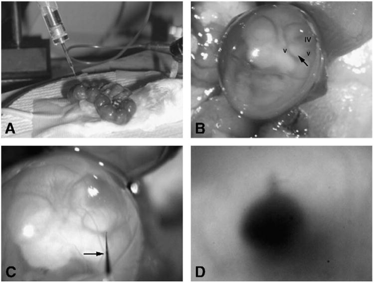
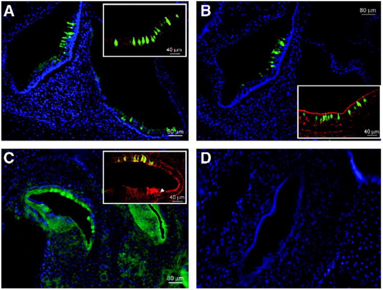
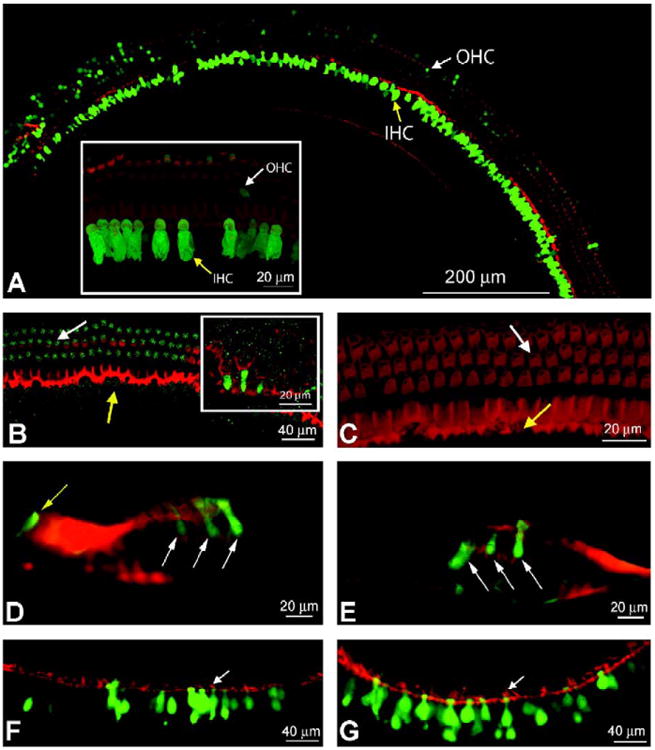
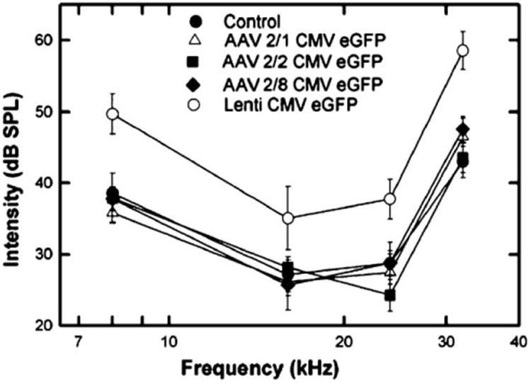
Congenital hearing deficits can be caused by a variety of genetic and acquired conditions. Complete reversal of deficits in the peripheral auditory system may require delivery of corrective genes to cochlear progenitor cells. We tested delivery of lentivirus and an array of recombinant adeno-associated viral (AAV) serotypes for efficiency and cellular specificity of transgene expression after in utero delivery to the developing mouse otocyst. Stability of expression and safety with respect to auditory function were then tested in those vectors that had the most favorable in utero cochlear transduction characteristics (AAV2/1, AAV2/8, and lentivirus). AAV2/1 was found to be the optimal vector for in utero cochlear gene transfer. It efficiently transduced progenitors giving rise to both inner and outer hair cells and supporting cells and had no adverse effect on cochlear cell differentiation. Further, it had no pathological effect on differentiated hair cells or the integrity of the auditory nerve or brain-stem nuclei as measured by auditory brain-stem response testing. AAV2/1 promises to be useful in further studies evaluating differentiation pathways of cochlear cells in health and disease and for developing gene-based therapies for congenital and acquired forms of peripheral hearing loss.


Figures

Fig. 1
Fig. 1
Methods of ex utero and trans-uterine microinjection. The surgical setup of the trans-uterine approach is shown as viewed through the dissecting microscope. (A) The uterus is exposed and the fetuses appear as a “string of beads” following midline laparotomy. (B) The left sagittal profile of the embryo at E12. Visible are the cardinal veins (v) and fourth ventricle (iv). The closed arrow points to the otocyst and the injection spot. (C) The microinjection pipette enters the amniotic sac (closed arrow). The light refracts the pipette tip as it enters the amniotic fluid. (D) After injection, dye is visible in the otocyst. Note the keyhole-shaped filled endolymphatic duct protruding away from the otocyst at the 12 o'clock position.
Fig. 2
Fig. 2
Sagittal cryosections of the murine cochlear and vestibular systems. eGFP-expressing hair cells and supporting cells are seen with fluorescence microscopy in cochlear and vestibular sagittal cryosections of P0 mice that had been injected at E12. (A) Vestibular section after exposure to AAV2/1 (original magnification 10×). The inset (original magnification 25×) shows transduced hair cells and supporting cells that express eGFP. Anti-myosin VIIa antibody is used as a hair cell marker and the reactive cells appear red. Hair cells that express eGFP as well as myosin VIIa appear green-yellow at the colocalization sites. (B) AAV2/8-injected cryosections are seen at 10× original magnification with a 25× original magnification inset. (C) Cochlear section after exposure to lentivirus and staining with an anti-eGFP antibody followed by FITC for better visualization. The white arrow in the inset indicates a myosin VIIa-stained hair cell. Nonspecific eGFP colocalization with this stain appears yellow-green. (D) Nuclei (stained blue with DAPI) outline the macula in the utricle in a sample from an AAV2/9-injected mouse. eGFP is absent.
Fig. 3
Fig. 3
Long-term gene expression in IHCs and OHCs. Confocal images of mature cochleae after in utero microsurgical viral injection. (A) A composite image of an apical turn is seen under confocal microscopy at original magnification 25×. Phalloidin, which stains F-actin in hair cells and supporting cells, appears red. A fragment of the cochlear turn is seen at original magnification 63× in the inset, which shows inner and outer hair cells that were transduced and express eGFP. Both images are of a cochlea injected with AAV2/1. White arrows mark OHCs and yellow arrows indicate transduced IHCs. (B) A segment of apical turn from a cochlea injected with lentivirus is seen at original magnification 25×. Three rows of OHCs are observed with a low level of eGFP expression. No IHCs are expressing eGFP. A 63× original magnification inset shows green fluorescent supporting cells indicating viral transduction. (C) A confocal projection image from an uninjected negative control at original magnification 63×. It is stained with phalloidin and exposed at a gain similar to that of the cochlear turn in (B) to demonstrate staining above background in (B). (D and E) AAV2/1- and 2/8-injected modiolar cross sections, respectively, at original magnification 40× are seen under light microscopy. The white arrows indicate infected OHCs and the yellow arrow indicates an infected IHC. (F and G) AAV2/1- and 2/8-injected utricular sections are seen at original magnification 20× under light microscopy. Many green fluorescent hair cells and supporting cells are observed. White arrows point to hair cell stereociliae as an indicator of hair cells. In several images, the cuticular plate can be distinguished by its yellow colabeling.
Fig. 4
Fig. 4
ABR responses in adult mice following in uterogene transfer. Average ABR thresholds in decibels referenced to sound pressure level (dB SPL) are plotted against frequency for groups of mice injected in utero with each viral vector. Frequencies tested were 8, 16, 24, and 32 kHz. Standard error of the mean ranges are plotted for each data point. A statistically significant threshold shift at each of the frequencies is seen between the lentiviral-injected mouse group and the uninjected controls (P < 0.001). No statistically significant difference between the rAAV-injected groups and controls was observed.

References

    1. Lalwani A. Development of in vivo gene therapy for hearing disorders: introduction of adeno-associated virus into the cochlea of the guinea pig. Gene Ther. 1996;3:588–592. - PubMed
    1. Zine A. Molecular mechanisms that regulate auditory hair-cell differentiation in the mammalian cochlea. Mol Neurobiol. 2003;27:223–237. - PubMed
    1. Izumikawa M, et al. Auditory hair cell replacement and hearing improvement by Atoh1 gene therapy in deaf animals. Nat Med. 2005;11:271–276. - PubMed
    1. Stone I, Lurie D, Kelley M, Poulsen D. Adeno-associated virus-mediated gene transfer to hair cells and support cells of the murine cochlea. Mol Ther. 2005;11:843–848. - PubMed
    1. Han J, et al. Transgene expression in the guinea pig cochlea mediated by a lentivirus-derived gene transfer vector. Hum Gene Ther. 1999;10:1867–1873. - PubMed

Publication types

MeSH terms

Substances