

TRPC1 protects human SH-SY5Y cells against salsolinol-induced cytotoxicity by inhibiting apoptosis
- PMID: 16765919
- PMCID: PMC2845452
- DOI: 10.1016/j.brainres.2006.04.104
TRPC1 protects human SH-SY5Y cells against salsolinol-induced cytotoxicity by inhibiting apoptosis
Abstract
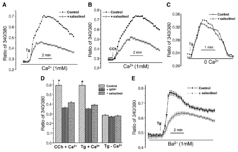
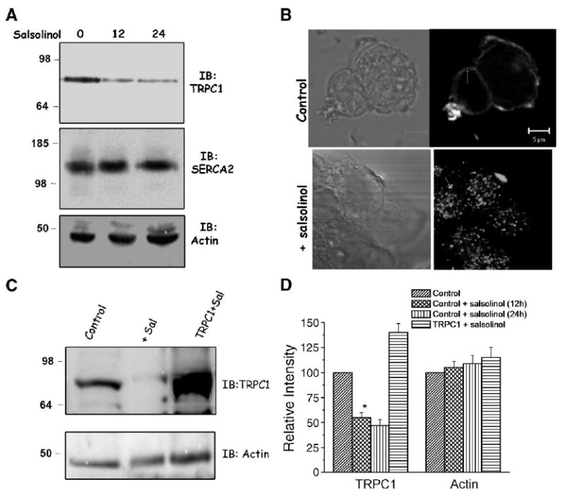
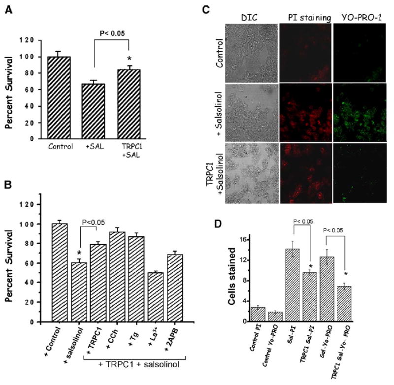
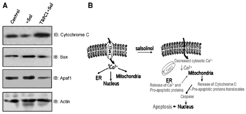
Salsolinol, an endogenous neurotoxin, may be involved in the pathogenesis of Parkinson's disease. In this study, we sought to determine whether salsolinol-induced cytotoxicity in SH-SY5Y human neuroblastoma cells, a cloned cell line which expresses dopaminergic activity, could be prevented by overexpressing a Ca(2+) channel, transient receptor potential (TRPC1) protein. Exposure of SH-SY5Y cells to 500 microM salsolinol for 12 h resulted in a significant decrease in thapsigargin or carbachol-mediated Ca(2+) influx. Consistent with these results, SH-SY5Y cells treated with salsolinol showed approximately 60% reduction in TRPC1 protein levels. Confocal microscopy also showed that SH-SY5Y cells treated with salsolinol had a significant decrease in the plasma membrane staining of the TRPC1 protein. Interestingly, overexpression of TRPC1 increases TRPC1 protein levels and also protected SH-SY5Y neuroblastoma cells against salsolinol-mediated cytotoxicity as determined by 3,[4,5-dimethylthiazol-2-yl]-2,5-diphenyltetrazolium bromide (MTT) assay. The protective effect of TRPC1 was blocked by the addition of TRPC1 blockers lanthanum, or 2APB. Activation of TRPC1 protein by either thapsigargin or carbachol further protected SH-SY5Y cells from salsolinol treatments. Staining of SH-SY5Y cells with an apoptotic marker (YO-PRO-1) showed that TRPC1 protein protects against apoptosis. Furthermore, TRPC1 overexpression also inhibited cytochrome c release and decreased BAX protein levels required for apoptosis. Taken together, these findings suggest that the reduction in cell surface TRPC1 protein expression in response to salsolinol may be a contributory factor in cellular toxicity of the dopaminergic neurons. Furthermore, overexpression of TRPC1 could inhibit apoptotic complex thereby increasing neuronal cell survivability in Parkinson's disease.
Figures
References
-
- Anglade P, Vyas S, Javoy-Agid F, Herrero MT, Michel PP, Marquez J, Mouatt-Prigent A, Ruberg M, Hirsch EC, Agid Y. Apoptosis and autophagy in nigral neurons of patients with Parkinson's disease. Histol Histopathol. 1997;12:25–31. - PubMed
-
- Baffy G, Miyashita T, Williamson JR, Reed JC. Apoptosis induced by withdrawal of interleukin-3 (IL-3) from an IL-3-dependent hematopoietic cell line is associated with repartitioning of intracellular calcium and is blocked by enforced Bcl-2 oncoprotein production. J Biol Chem. 1993;268:6511–6519. - PubMed
-
- Bellomo G, Perotti M, Taddei F, Mirabelli F, Finardi G, Nicotera P, Orrenius S. Tumor necrosis factor alpha induces apoptosis in mammary adenocarcinoma cells by an increase in intranuclear free Ca2+ concentration and DNA fragmentation. Cancer Res. 1992;52:1342–1346. - PubMed
-
- Bembenek ME, Abell CW, Chrisey LA, Rozwadowska MD, Gessner AW, Brossi A. Inhibition of monoamine oxidase-A and oxidase-B by simple isoquinoline alkaloids-racemic and optically active 1,2,3,4-tetrahydroisoquinoline, 3,4-dihydroisoquinoline, and fully aromatic isoquinoline. J Med Chem. 1983;33:147–152. - PubMed
-
- Berridge MJ, Lipp P, Bootman MD. The versatility and universality of calcium signalling. Nat Rev, Mol Cell Biol. 2000;1:11–21. - PubMed
Publication types
MeSH terms
Substances
Grants and funding
LinkOut - more resources
Full Text Sources
Medical
Molecular Biology Databases
Research Materials
Miscellaneous
